Download
1 / 45
450 likes | 684 Views
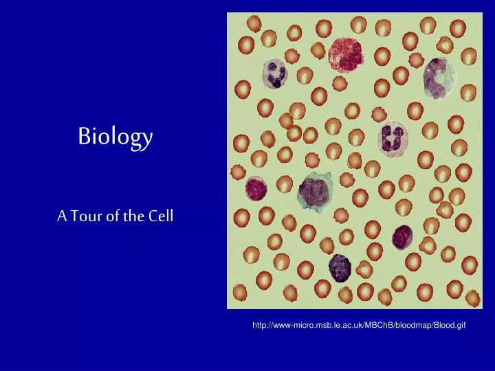
Biology. A Tour of the Cell. http://www-micro.msb.le.ac.uk/MBChB/bloodmap/Blood.gif. A cell is the smallest unit of life . They can vary in size, shape and function (structure determines function). The light microscope led the way to knowledge of the cell . Microscopy.

E N D
Biology A Tour of the Cell http://www-micro.msb.le.ac.uk/MBChB/bloodmap/Blood.gif
A cell is the smallest unit of life. They can vary in size, shape and function (structure determines function). Thelight microscope led the way to knowledge of the cell. Microscopy http://www.microscope-microscope.org/basic/microscope-images/138-microscopes-lg.jpg
Microscopes: • A light microscopemagnifies objects (specimens) ~1000x their size. Most cellular structures CANNOT be seen. • You will work with a light microscope in the lab. http://www.dsbn.edu.on.ca/schools/Westlane/Science/simon/SBI3C1/micro.gif
Electron microscopesgive more detail and magnify a million times the object’s size. • However, the organism dies when using an electron microscope. There are 2 types of electron microscopes: • A scanning electron microscope (SEM)gives a 3D image of a specimen/object. • A transmission electron microscope (TEM)transmits electrons to view the interior of an object.
How the Scanning Electron Microscope works: SEM http://w3.salemstate.edu/~pkelly/sem/image003.jpg
TEM How the Transmission Electron Microscope works:
TEM SEM http://img72.imageshack.us/img72/2392/071003100546198ddedh5.jpg http://www.st-andrews.ac.uk/~dclogan/Fig3.jpg
A history lesson: • Robert Hooke (1665) named the cell after looking at cork under the microscope. http://www.google.com/imgres
Anton van Leeuwenhoek (LAY-von-hohk) (1670’s) developed a simple light microscope &discovered unicellular organisms and called them “little beasties”. • Rudolf Virchow (1855) stated that all cells came from pre-existing cells (a.k.a.Cell Theory). http://www.google.com/imgres
The Cell Theory (Virchow) has 3 basic principles: • Cells are the basic units of life. • All organisms are made of 1 or more cells. • All cells arise from existing cells. http://www.leksikon.org/images/virchow_rudolf.jpg http://www.dmturner.org/Teacher/Pictures/Cell%20reproduction.jpg
Cell Structures: • All cells have an enclosure called a cell, or plasma membrane. • This functions as the gate keeper and controls what enters and exits the cell. • It is mainly composed of phospholipids and proteins. http://www.google.com/imgres
Within the cell is thecytoplasm. This is a semifluid substance that contains the organelles. • The organellesare small structures that have specific functions within the cells. http://www.google.com/imgres
The cytoskeletonis a protein network within the cytoplasm that helps support the cell and helps the cell maintain or change its shape. It also • Anchors organelles • Enables the cell to move • Allows materials to move throughout the cell • Composed ofmicrotubules & microfilaments http://www.google.com/imgres
The nucleusis the control center of the cell • It is surrounded by the nuclear envelope • It contains thechromosomes. There are 46 chromosomes in the human nucleus (in every cell of the human body). The chromosomesare the DNA (genetic material). • The nucleus is only found in eukaryotic cells! http://www.google.com/imgres
The nucleuscontains thenucleolus (if more than 1, nucleoli). • Nucleolus: makes ribosomes • Ribosomes make proteins. http://www.google.com/imgres
Prokaryotic Cells Bacteria NO organelles(membrane-bound structures) Containedw/in cell membrane & cell wall, contain ribosomes, 1 circular chromosome innucleoid region(NO nucleus)& plasmids (extra pieces of DNA) Eukaryotic Cells Protists, Fungi, Plants, & Animals Contained w/in cell membrane (may have a cell wall) Contain nucleus & other membrane-bound organelles Means ‘true kernel’ 2 Types of Cells
Prokaryotes: These are bacteria (in Kingdom Monera). They are unicellular organisms. These were the first cells. They are very small cells and are very simple cells. There are 2 types of cells: http://www.singleton-associates.org/gifs/cell.jpg
2. Eukaryotes:found in all other kingdoms except Monera. • These have a nucleus (as well as a cell membrane and the majority of the organelles being discussed, depending on the type of organism). http://www.google.com/imgres
http://micro.magnet.fsu.edu/cells/plants/images/plantcell.jpghttp://micro.magnet.fsu.edu/cells/plants/images/plantcell.jpg
Ribosomesmake proteins from amino acids; can be found suspended within the cytoplasm or attached to the endoplasmic reticulum. (not really organelles; these are cellular components) • The endoplasmic reticulum (ER) is a network of membrane that produces materials for the cell. There are 2 types: • The rough ERcontains ribosomes and functions in protein synthesis and makes new cell membrane. • The smooth ERmakes lipids, process carbohydrates and breaks down toxins.
The golgi apparatus is composed of flat membranous save that modify, package and distribute molecules (warehouse of the cell). http://www.google.com/imgres
Vacuole • Vacuolesare membrane-bound organelles that have various functions. • Some store food, water, proteins, ions, or wastes. Generally these are large and centralized. • Lysosomescontain digestive enzymes that break down large molecules and old organelles that the cell no longer needs. http://www.google.com/imgres
Chloroplasts, double-membrane-bound organelles, perform photosynthesis. • This is the process of making sugar (synthesis) in the presence of light (photo). • Plants (some bacteria & protists) make their own food (a.k.a.autotrophic). http://www.daviddarling.info/images/chloroplast.jpg
The mitochondriais the “powerhouse of the cell” b/c it changes stored enter from food into useable chemical energy (ATP) for chemical reactions. • ATP = adenosine triphosphate(energy ‘currency’ of cells) • The chemical reactions are cellular respiration. http://scienceblogs.com/worldsfair/Mitochondria.jpg
Cellular Structures • Ciliaare short hair-like projections that are in the surface on the cell usually in large number. (NOT organelles) • Beat in unison and aid in the cell’s movement or in the movement of fluid over the cell. http://www.google.com/imgres
Flagellaare long tail-like projections that are on the surface of the cell. (NOT organelles) • Usually 1 to 3 of these. • In prokaryotic cells, they spin like propellers. • In eukaryotic cells, they move like whips. http://www.google.com/imgres
Cell Wall Plants differ from animal cells. In plants: • A cell wallsurrounds the cell membrane. • The cell wallis a rigid outer covering that protects and maintains the shape of the plant cell. • Fungi, algae (a type of protist) and bacteria also have cell walls but the composition is different. http://www.google.com/imgres
Plants LACK lysosomes. • Animals are heterotrophic, meaning they must consume food. Animal cells: • LACK a cell wall but have cytoskeletons for structural support. • LACK chloroplasts • Contain small vacuoles (instead of a large centralized one) • Have lysosomes
BOTH Animal and Plant Cells Contain: • Organelles previously mentioned (nucleus, ER, mitochondria, ER, ribosomes, etc) • Cell membranes • DNA (in chromosomes)
Cell/plasma membraneis composed of aphospholipidbilayer (2 layers of phospholipids) with proteins interspersed. Phospholipids have a hydrophilic head & hydrophobic tail Fluid & flexible SEMI-PERMEABLE MEMBRANE http://www.bioteach.ubc.ca/Bio-industry/Inex/graphics/phospholipid.gif
Cell Membrane http://www.hallym.ac.kr/~de1610/histology/cell-3.jpg
Membrane Functions • Cell membranes are semipermeable. This means that some things pass through the membrane while others cannot pass through (this depends on the size & charge of the molecule). • Passive transport is the movement of a substance across a membrane without energy input. • Active transport is the movement of a substance across a membrane with the input of energy.
Concentration=[ ]. • Molecules move from a higher [ ] gradient to a lower [ ] gradient. • A [ ] gradient is the difference between the [ ] of a particular molecule in 1 area and its [ ] in an adjacent area. • The rate of diffusiondepends on temperature and size of molecules involved (molecules move faster at higher temperatures and smaller molecules move faster than larger molecules). • Once molecules are dispersed evenly, equilibriumis reached and diffusion stops.
Passive Transport • Diffusion is the movement of molecules from a higher concentration to a lower concentration. http://www.google.com/imgres
Facilitated diffusion is the diffusion of molecules with the help of a carrier protein embedded within a cell membrane. http://www.google.com/imgres
Osmosis is the diffusion of water. • Diffusion, facilitated diffusion and osmosis are all type of passive transport. These do NOT require energy (occur spontaneously). http://www.biologycorner.com/resources/osmosis.jpg
Active transport in cells usually occurs with the help of carrier proteins but REQUIRE energy. An example is the sodium-potassium pump (Na+/K+ pump). http://web.ahc.umn.edu/~mwd/cell_www/images/Na-Kpump.png
When comparing 2 solutions with a membrane between them, there are 3 types of solutions: • Hypertonic: the fluid outside a cell has a higher [solute] than the cytoplasm inside the cell. In this case, water diffuses out of the cell. • Isotonic: the [solute] outside the cell= the [solute] inside the cell. In this case, no osmosis will occur. • Hypotonic: the fluid outside a cell has a lower [solute] than the cytoplasm inside the cell. In this case, water will move inside the cell.
Bulk Transport: 1. Exocytosis: exo=exit; cyto=cell • Wastes and cell products are packaged in vesicles by the golgi apparatus. • The vesicles fuse with the cell membrane and leave the cell http://www.octc.kctcs.edu/gcaplan/anat/images/Image152.gif
2. Endocytosis: endo=within; cyto=cell • A portion of the cell membrane surrounds a substance outside of the cell & pinches off to form a vesicle • The vesicle moves inward and fuses with other organelles • This includes • Pinocytosis:cell drinking • Phagocytosis:cell eating
http://www.gla.ac.uk/~jmb17n/Teaching/L2teaching/Agpres/Figures/Endocytosis.jpghttp://www.gla.ac.uk/~jmb17n/Teaching/L2teaching/Agpres/Figures/Endocytosis.jpg
To review cell structures and reproduction, click on these links and do some activities: http://www.cellsalive.com/cells/cell_model.htm http://biolabs.wikispaces.com/Cell+Drawings