Download
1 / 25
250 likes | 350 Views
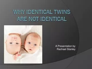
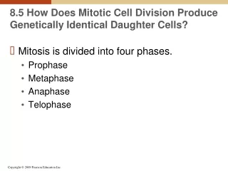
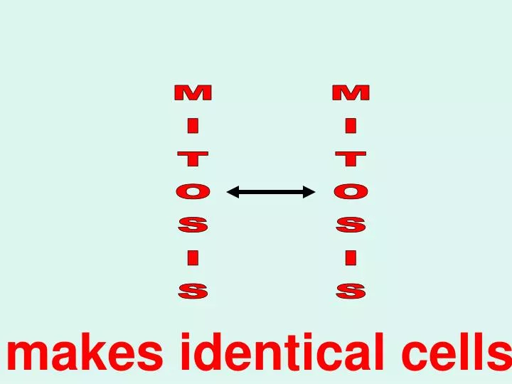
MITOSIS. MITOSIS. makes identical cells. Control of Cell Division and Cell Death. Human cells appear to be programmed to undergo only so many cell divisions About 50 in cell cultures. Fingers and toes form from these paddlelike hands and feet.

E N D
MITOSIS MITOSIS makes identical cells
Control of Cell Division and Cell Death • Human cells appear to be programmed to undergo only so many cell divisions • About 50 in cell cultures Fingers and toes form from these paddlelike hands and feet • During fetal development, many cells are programmed to die, apoptosis Apoptosis happens if cells of the human myeloid cell line are deprived of growth factors. SEM 10,000. Only cancer cells and stem cells can divide endlessly
Controlling the Cell Cycle • The eukaryotic cell cycle is controlled by feedback at three checkpoints • 1. Cell growth is assessed at the G1 checkpoint • 2. DNA replication is assessed at the G2 checkpoint • 3. Mitosis is assessed at the M checkpoint • Cancer is characterized by cells that have lost the ability to control their division see Figure 8.9 G0 is an extended rest period
What is Cancer? • Cancer is unrestrained cell growth and division • Most cancers result from mutations in growth-regulating genes • There are two general classes of these genes • 1. Proto-oncogenes • Encode proteins that simulate cell division • If mutated, they become oncogenes • 2. Tumor-suppressor genes • Encode proteins that inhibit cell division • Can undergo metastasis • Leave the tumor and spread throughout the body
Cancer can be caused by chemicals, radiation or viruses • Rous Sarcoma Virus (RSV) chicken cancer from cell-free extracts • - Peyton Rous 1911 Nobel Prize 1966 • - Oncogenes - cancer causing genes • - Protooncogenes- normal genes which stimulate cell growth • - growth factors, receptors, • signal transduction proteins, etc
The p53gene plays a key role in the G1 checkpoint of cell division The p53 protein (the gene’s product), monitors the integrity of DNA If DNA is damaged, the protein halts cell division and stimulates repair enzymes If the p53 gene is mutated Cancerous cells repeatedly divide No stopping at the G1 checkpoint Cancer and Control of the Cell Cycle
New molecular therapies for cancer Receiving the signal to divide Stopping tumor growth Stepping on the gas Passing the signal via a relay switch Amplifying the signal Releasing the “brake” Checking that everything is ready
Prokaryotes Have a Simple Cell Cycle • Prokaryotic cells divide asexually • These cells possess a single • circular chromosome, containing • about 1000 genes • The chromosome is replicated • The cell then divides into two cells, • a process called binary fission • – 17 million daughter cells in 8-hours
Cell division in eukaryotes is more complex than in prokaryotes because 1. Eukaryotic contain far more DNA 2. Eukaryotic DNA is packaged differently It is in linear chromosomes compacted with proteins Eukaryotes Have a Complex Cell Cycle • Some eukaryotes also • make exact copies of • themselves via asexual • reproduction amoeba
The Cell Cycle Interphase • G1 phase • Primary growth phase • S phase • DNA replication • G2 phase • Microtubule synthesis • M phase (MITOSIS) • Chromosomes pull apart • C phase (CYTOKINESIS) • Cytoplasm divides Figure 8.5
Chromosomes • The DNA helix is wrapped around positively-charged proteins, called histones • ~200 nucleotides of DNA + histones= nucleosome • During interphase, the DNA is coiled into chromatin but not visible chromosomes • A chromosome is a complex of one molecule of DNA (~ 40%) • and proteins (~ 60%) • A typical human chromosome contains about 1000 genes • ~ 140 million nucleotides in its DNA, ~5 cm in stretched length ^Figure 8.2
Homologous Chromosomes • Diploid cellshave two SIMILAR* chromosomes • called homologous chromosomesorhomologs • Humans have 46 chromosomes or 23 pairs of homologs(similarchromosomes, one from this person’s Mom and one from their Dad) Figure 8.4 The chromosomes can be organized by size into a karyotype * Important
Sister Chromatids • Replicated chromosomes consist of twoIDENTICAL* • sister chromatids held together at the centromere • DNA replication occurs in • S phase(Sister) • Homologsare SIMILAR* chromosomes * Important see Tables 8.1 & 8.2 for terms
Before a cell starts dividing, the chromosomes duplicate • Because the strands of the double helix are complementary A+T C+G an exact copy of the DNA can be made • An emergent property
Chromosomes duplication (DNA replication or Synthesis) produces IDENTICAL sister chromatids • When the cell divides, • the sister chromatids separate • Each of the two daughter cells has a • complete and identical set of chromosomes
Eukaryotic cells divide in one of two ways Mitosis Occurs in somatic (non-reproductive) cells Meiosis Occurs in germ (reproductive) cells Results in the production of gametes Haploid gametes (n = 23) Egg cell Sperm cell MEIOSIS FERTILIZATION Diploidzygote (2n = 46) Multicellulardiploid adults (2n = 46) Mitosis anddevelopment MITOSIS makes identical diploid (2n) cellsMEIOSIS makes haploid (1 n) gametes
Mitotic Cell Cycle • M phase MITOSIS • Chromosomes pull apart • Prophase • Metaphase • Anaphase • Telophase • C phase CYTOKENESIS • Cytoplasm divides • INTERPHASE • G1 phase • Primary Growth phase • S phase • DNA Synthesis • G2 phase • Growth & Microtubule synthesis Figure 8.5 Chromosomes are decondensed during interphase chromatin
Prophase • Chromosomes coil • Nuclear membrane breaks down • Spindle fibers form see Figure 8.7
see Figure 8.7 Metaphase • Chromosomes line up on the midline • Spindle fibers attach to centromeres
Anaphase • Centromeres divide • Spindle fibers shorten • Sister chromatids separate and move to opposite poles see Figure 8.7
Telophase • Cell elongates • Nuclear membrane reforms • Chromosomes uncoil • Spindle disappears see Figure 8.7
Cytokinesis • Division of the cytoplasm • Cleave furrow forms at equator of the cell • Constriction tightens by contraction of filaments • Cell is divided into two identical cells see Figure 8.7
see Figure 8.8 • Animal cells • Plant cells • Cleavage furrowforms, pinching the cell in two • Cell plateforms, dividing the cell in two • Cytokinesis
skin Deadcells - Asexual reproduction, development, growth and cell replacement are mitotic divisions Epidermis, the outer layer of the skin Dividingcells Dermis