Download
1 / 25
280 likes | 1.11k Views
Karyotyping. Preparing human chromosomes for observation. History of Karyotyping. 1882: Flemming saw human chromosomes dividing in skin cells. 1956. Human chromosome number confirmed at 46 by J.H. Tjio and Albert Levan. 1958.

E N D
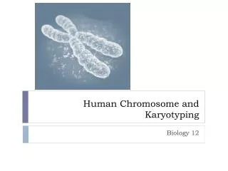
Karyotyping Preparing human chromosomes for observation
History of Karyotyping • 1882: Flemming saw human chromosomes dividing in skin cells
1956 • Human chromosome number confirmed at 46 by J.H. Tjio and Albert Levan.
1958 • Lejuene discovered that Down syndrome is caused by a diploid number of 47 (Trisomy 21)
1960 • Technique developed for growng human leukocytes in cell culture. They grow much faster than skin cells.
HOW TO KARYOTYPE • 1. Obtain living cells • Draw a blood sample to get leukocytes • Or • Obtain amniotic fluid by amniocentesis • Or • Obtain fetal cells by chorion villus sampling
How to Karyotype • Stimulate cell division by placing cells to culture medium in a Petri dish and adding phytohemaglutinin.
How to Karyotype • Add colchicine to cells to inhibit production of spindle fibers. • Cell division stops at metaphase.
How to Karyotype • Treat cells with 1% saline solution. • This hypotonic solution causes cells to swell up. • Chromosomes can spread out inside cells.
How to Karyotype • Hold cells 3 to 4 feet above a microscope slide and drop them onto a glass slide • SPLAT!!!! • Dry the cells so they STICK to the glass
How to Karyotype • STAIN cells so the chromosomes are visible • Many staining options exist: • C band is easiest • G & R banding • Q banding • FISH
Chromosome staining • 1. C Band: use Giemsa, a dark blue and purple stain • 2. G & R banding uses trypsin enzyme to digest away chromosome proteins, then stain to show BANDING patterns • 3. Q banding: use quinicrine mustard, a fluorescent dye, to stain cells….they glow, but the stain is dangerous.
FISH staining • Fluorescence In-Situ Hybridization • A segment of DNA from each chromosome is bonded to a different color dye. • These DNA segments are added to the cells in each Petri dish. • Matching DNA segments STICK to the correct chromosome which then glows a certain color! SWEET!
Mary Lyon • Why should females have two X chromosomes while healthy males only have one? • The Lyon Hypothesis: Only one X chromosome is used by your cell. The other one is ignored. • What evidence should we look for?
The Barr Body • Robert Barr spotted these blobs of chromatin coiled up inside the cell nuclei of females.
Barr body is caused by X-inactivation • As a female embryo develops, one X chromosome in each cell turns into a Barr body and is not used. • If a woman is homozygous, it causes no changes. • If a woman is heterozygous, some of her cells will look one way, while other cells look a different way.
Example of X-inactivation • Calico cat: a mosaic trait
Example of X-inactivation • Tortoiseshell cat: a mosaic trait
Before the cells arrived • HeLa cells grown in cell culture • Cells treated with colchicine (microtubule inhibitor) • Cells rinsed and centrifuged • Cells placed in hypotonic solution, with distilled water, acetic acid and alcohol preservative.
Today’s preparation • Place wet glass slide at 45 deg. Angle • Gently resuspend cells with pipette • Hold pipette 24 inches above slide. • Allow ONE DROP of cell suspension to “SPLAT” onto the slide about 3 cm from the upper end, and tumble down the slide.
Part 2 • Carefully apply 6 to 8 drops from various heights, ONE DROP at a time • Gently blow across slide to dry the cell suspension. • AIR DRY the slides completely.
Staining • Dip the slide into the STAIN 1 tube for 1 second only • Dip the slide into the STAIN 1 tube again for 1 second only • Dip the slide into the STAIN 1 tube again for 1 second only • Drain the stain from the slide
Staining • Dip the slide into the STAIN 2 tube for 1 second only • Dip the slide into the STAIN 2 tube again for 1 second only • Dip the slide into the STAIN 2 tube again for 1 second only • RINSE with a slow stream of water • AIR DRY