Download
1 / 4
40 likes | 159 Views
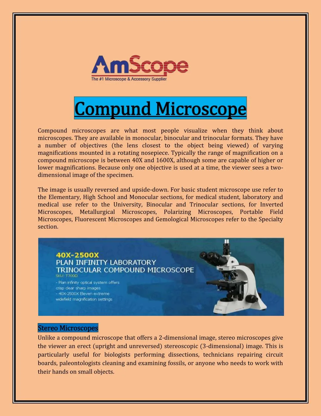
Compound microscopes are what most people visualize when they think about microscopes. They are available in monocular, binocular and trinocular formats. They have a number of objectives (the lens closest to the object being viewed) of varying magnifications mounted in a rotating nosepiece.<br>

E N D
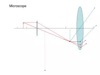
Compund Compund Microscope Microscope Compound microscopes are what most people visualize when they think about microscopes. They are available in monocular, binocular and trinocular formats. They have a number of objectives (the lens closest to the object being viewed) of varying magnifications mounted in a rotating nosepiece. Typically the range of magnification on a compound microscope is between 40X and 1600X, although some are capable of higher or lower magnifications. Because only one objective is used at a time, the viewer sees a two- dimensional image of the specimen. The image is usually reversed and upside-down. For basic student microscope use refer to the Elementary, High School and Monocular sections, for medical student, laboratory and medical use refer to the University, Binocular and Trinocular sections, for Inverted Microscopes, Metallurgical Microscopes, Polarizing Microscopes, Portable Field Microscopes, Fluorescent Microscopes and Gemological Microscopes refer to the Specialty section. Stereo Stereo Microscopes Microscopes Unlike a compound microscope that offers a 2-dimensional image, stereo microscopes give the viewer an erect (upright and unreversed) stereoscopic (3-dimensional) image. This is particularly useful for biologists performing dissections, technicians repairing circuit boards, paleontologists cleaning and examining fossils, or anyone who needs to work with their hands on small objects.
Most stereo microscopes are used at magnifications from 5X to 90X, but with the proper microscope and accessories, magnifications up to approaching 180X can be achieved. For educational and simple hobby use refer to the Binocular Stereo Microscopes. To customize and build a stereo microscopy system for industrial or advanced applications refer to the Trinocular, Boom and Inspection Microscopes. These types of system will allow one to customize stands and illumination systems for any type of application. For Research Grade Stereo Microscopes we feature the zoom and boom Series. These systems are designed for critical viewing for research, medical, forensic and other high end stereo uses. Microphotography For many applications the ability to capture, display, and preserve specimen images is of equal or greater importance than actually viewing the specimen through the eyepieces. Photomicrography (35mm and other chemical formats) has been a common option on microscopes for decades, but the recent development of relatively inexpensive CCD (charged couple device) video and digital cameras has greatly increased both the popularity and flexibility of microscope imaging. Instead of clicking through slides during a lecture, university professors can now display real-time video images on projection televisions; petroleum geologists can e-mail images of core samples to their laboratories from remote locations around the world; oncologists can refer to CD or on-line catalogues of cell images to help them make faster and more accurate diagnoses. For video and digital imaging, refer to “CCD, USB, Camera”, LP Digital Scope and HP Digital Scope sections. There are many different methods for capturing, displaying, and recording microscope images, and each has its own advantages and disadvantages. It would be impossible to cover all of these options here but one basic piece of information will be important in selecting your microscope: While it is possible to mount a camera on a monocular or binocular microscope (note: a binocular microscope has two eyepieces, but is not necessarily a stereo microscope), it is far better to use a trinocular microscope designed for camera work. Trinocular models have two eyepieces for normal viewing, plus a third "phototube" on which you can mount a camera without interfering with the normal operation of the microscope. Trinocular microscopes are ideal for photo, digital or video applications. Remember, depending upon your application additional components are required on your microscope depending upon your use. Our friendly sales force can assist you in choosing the correct items required.
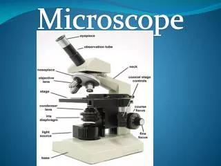
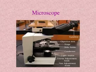
How How To To Use Use A A Properly Properly Use Use A A Microscope Microscope 1. When transporting the microscope, carry it close to your body with one hand on the arm and the other on the base. If you are unsure what part of the microscope is which, I recommend starting at our Microscope Parts & Functions section, found here. 2. Once you have removed the dust cover from the microscope if using one, and have plugged the microscope in, arrange the excess cord in a manner that will prevent you from tripping on it and knocking the microscope over. A microscope is a precision instrument, so any sharp movements or impact can set the fine elements of the unit off. 3. As a general rule, always start and end with the lowest power objective on the microscope (usually 4X), as it is easiest to focus and center the sample in the image on a lower power. Also, since it is the shortest objective lens, there is the least chance of scratching the lens when situating or removing the slide from the microscope. The below image shows how to properly mount the sample onto your silde. 4. Turn on the microscope and place the slide on the microscope stage with the specimen directly over the circle of light. Doing this will give you a 90% chance of finding the specimen as soon as you look through the eyepiece. If your microscope is monocular (it has only one eyepiece), close or cover your other eye for accuracy and comfort when viewing. If your microscope is binocular, adjust the interpupillary distance (the distance between the eyepieces) by either sliding or rotating the eyepieces (called adjusting the diopter if present on the microscope) appropriately until you can see only one circle of light with both eyes open. 5. If you are on the lowest magnification level, focus the image by first turning the coarse focus knob. If you can’t get it at all into focus using the coarse knob, then switch to the fine focus knob. 6. Adjust the diaphragm as you look through the eyepiece. You will begin to notice that more detail is visible when you allow in less light. Too much light tends to give the specimen a washed-out appearance. 7. Once you have focused the specimen on low power (usually a 4X objective), center the specimen in your field of view, then, without changing the focus knobs, switch it to a higher magnification objective (10X objective, then 40X objective). If you don’t center the specimen you will lose it when you switch to higher powers. History of the Microscope The Beginning The Beginning During that historic period known as the Renaissance, after the "dark" Middle Ages, there occurred the inventions of printing, gunpowder and the mariner's compass, followed by the discovery of America.
Equally remarkable was the invention of the light microscope: an instrument that enables the human eye, by means of a lens or combinations of lenses, to observe enlarged images of tiny objects. It made visible the fascinating details of worlds within worlds. Invention Invention of of Glass Long before, in the hazy unrecorded past, someone picked up a piece of transparent crystal thicker in the middle than at the edges, looked through it, and discovered that it made things look larger. Someone also found that such a crystal would focus the sun's rays and set fire to a piece of parchment or cloth. Magnifiers and "burning glasses" or "magnifying glasses" are mentioned in the writings of Seneca and Pliny the Elder, Roman philosophers during the first century A. D., but apparently they were not used much until the invention of spectacles, toward the end of the 13th century. Glass Lenses Lenses They were named lenses because they are shaped like the seeds of a lentil. The earliest simple microscope was merely a tube with a plate for the object at one end and, at the other, a lens which gave a magnification less than ten diameters -- ten times the actual size. These excited general wonder when used to view fleas or tiny creeping things and so were dubbed "flea glasses." Birth Birth of of the About 1590, two Dutch spectacle makers, Zaccharias Janssen and his son Hans, while experimenting with several lenses in a tube, discovered that nearby objects appeared greatly enlarged. That was the forerunner of the compound microscope and of the telescope. In 1609, Galileo, father of modern physics and astronomy, heard of these early experiments, worked out the principles of lenses, and made a much better instrument with a focusing device. The father of microscopy, Anton van Leeuwenhoek of Holland (1632- 1723), started as an apprentice in a dry goods store where magnifying glasses were used to count the threads in cloth. He taught himself new methods for grinding and polishing tiny lenses of great curvature which gave magnifications up to 270 diameters, the finest known at that time. These led to the building of his microscopes and the biological discoveries for which he is famous. He was the first to see and describe bacteria, yeast plants, the teeming life in a drop of water, and the circulation of blood corpuscles in capillaries. During a long life he used his lenses to make pioneer studies on an extraordinary variety of things, both living and non living, and reported his findings in over a hundred letters to the Royal Society of England and the French Academy. For more information please visit For more information please visit the Light Light Mi Microscope croscope https://www.amscope.com https://www.amscope.com