Download
1 / 6
60 likes | 63 Views
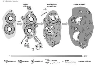
This study examines the mechanisms of cellular infiltration, myofibroblast accumulation, and tubular atrophy in tubulointerstitial fibrosis. The role of extracellular matrix (ECM) infiltration, epithelial mesenchymal transition (EMT), apoptosis, and proliferation is investigated. Additionally, the involvement of growth factors/cytokines, chemokines, and the renin-angiotensin system in fibrosis progression is explored.

E N D
Fig 1, Bascands & Schanstra cellular infiltration myofibroblast accumulation tubular atrophy ECM infiltration UUO EMT apoptosis proliferation proliferation - + ECM Collagen production fibroblast growth factors/cytokines epithelial cell macrophage apoptotic cell myofibroblast
Fig 2, Bascands & Schanstra CCR1 CD44 circulation endothelial cells L-selectin chemokines osteopontin sulfatide interstitium
Fig 3, Bascands & Schanstra TGF Interstitial fibroblast activation Epithelial Mesenchymal Transition 1) Loss of epithelial adhesion properties 2) a-SMA expression and actin reorganization 3) Disruption of the tubular basement membrane 4) Enhances epithelial cell migration and invasion tubule fibroblast myofibroblast MMP9 expression Bone marrow derived cells matrix accumulation tPA knockout interstitium
Fig 4, Bascands & Schanstra matrix PAI-1 MMP plasmin PA angiotensinogen kininogen renin ACE-inhibitors kallikrein AI bradykinin TGF AII ACE AT1 receptor inactive peptides cellular infiltration HGF plasminogen B2 receptor pro-MMP matrix MMP-9 EMT
Fig 5, Bascands & Schanstra P IB IB NF-B NF-B NF-B angiotensinogen renin AI ACE AII TNFa postive regulation loops AT1 AT2 TNFR1 TNFR2 IB kinases TNFa angiotensinogen gene transcription inflammation matrix expression proinflammatory and profibrotic cytokines IkB degradation NF-kB motif Nucleus fibrosis
Fig 6, Bascands & Schanstra R-I R-I R-II R-II TGF TGF TGF P SMAD2/3 P SMAD2/3 P SMAD4 decorin R-I SMAD2/3 R-II RhoA/Rock stress fiber formation p38MAPKs reorganization of actin cytoskeleton plasmin integrin aVb6 SMAD4 Nucleus gene transcription EMT cellular infiltrationapoptosis enhancer of TGFb regulated genes latent-TGF Tubulointerstitial fibrosis