Download
1 / 29
380 likes | 789 Views
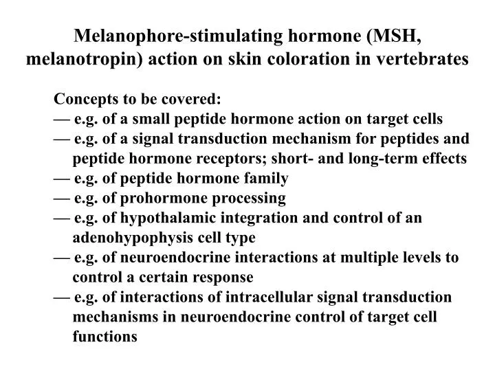
Melanophore-stimulating hormone (MSH, melanotropin) action on skin coloration in vertebrates. Concepts to be covered: — e.g. of a small peptide hormone action on target cells — e.g. of a signal transduction mechanism for peptides and peptide hormone receptors; short- and long-term effects

E N D
Melanophore-stimulating hormone (MSH, melanotropin) action on skin coloration in vertebrates Concepts to be covered: — e.g. of a small peptide hormone action on target cells — e.g. of a signal transduction mechanism for peptides and peptide hormone receptors; short- and long-term effects — e.g. of peptide hormone family — e.g. of prohormone processing — e.g. of hypothalamic integration and control of an adenohypophysis cell type — e.g. of neuroendocrine interactions at multiple levels to control a certain response — e.g. of interactions of intracellular signal transduction mechanisms in neuroendocrine control of target cell functions
Why colour change? Significance? — mimicking background — skin heat absorption and reflection — visual signalling cues (behaviour) — protection from UV radiation
Two types of colour change: Morphological Colour Change —synthesis of pigments and increase in number of pigmented cells (chromatocytes and chromatophores) — slow, long-term effects • Physiological Colour Change • — redistribution of pigments inside chromatophores • — pigments contained in vesicular structures called chromatosomes • — relatively fast, short-term effects
Pro-opiomelanocortin (POMC) (precursor for multiple potentially active peptides) gamma alpha beta CLIP END MSH MSH MSH ACTH beta-LPH gamma-LPH Processing at ACTH beta-LPH PARS DISTALIS Processing at alpha CLIP gamma-LPH END MSH PARS INTERMEDIA MSH - Melanophore stimulating hormone, melanotropin ACTH - Adrenocorticorticotropic hormone, corticotropin LPH - Lipotropic Hormone, lipotropin END - Endorphin CLIP - Corticotropin-like peptide (probably inactive)
Melanosome (organelles) Melanohpore (cell) epidermis Basal Lamina Other chromatophores irridophore dermis melanophore Dermal and epidermal chromatophore units in amphibian skin
Under MSH action: melanosomes dispersed melanophore epidermis Basal Lamina irridophore dermis melanophore Dermal and epidermal chromatophore units in amphibian skin responding to MSH in physiological colour change
Neuroendocrine integration of physiological colour change (1) Relative thickness of arrows of hypothalamic & pituitary influences indicate relative signal strength
Neuroendocrine integration of physiological colour change (2) Relative thickness of arrows of hypothalamic & pituitary influences indicate relative signal strength
For proper regulation of physiological colour change, neuro-endocrine interaction must occur at multiple levels: — Sensory input concerning light:dark information — Integration at the level of the brain — Control of hypothalamic neurosecretion — Control of pituitary endocrine secretion — Control of neuronal innervation of the skin — Integration of endocrine and neuronal action at the skin
How is specificity of hormone action maintained if hormones travel via the blood stream of tissue fluid? — target cells have the appropriate receptors for the appropriate hormone — receptors are molecules that recognize the hormone in a specific manner (i.e., only the appropriate molecules will bind the receptor - usually only a few very closely related molecules of similar molecular shape will bind) — hormone binds to these receptors and can activate these receptors — (note: not all molecules that can bind a receptor will activate the receptor - concept of receptor agonists and antagonists)
How is specificity of hormone action maintained if hormones travel via the blood stream of tissue fluid? (continued) — activation of these receptors by agonists alter biochemical events in the cell which cause appropriate responses to be generated — processes involved in the linking of receptor activation to the appropriate cellular response are called signal transduction events — intracellular chemical molecules involved in these signal transduction events are generally called “second messengers” (i.e., hormone is the first chemical messenger and these intracellular chemical signalling molecules are the second chemical messengers in the sequence of event)
Basic Elements in Peptide Hormone Signal Transduction at the Target Cell Hormone Extracellular primary signal Receptor Signal recognition site G-protein Event coupler and transducer Generation and/or modulation of second messengers levels Generation of internal signals Modulation of some intracellular biochemical event Changes in intracellular events Biological response Modification of output
N outside I II III IV V VI VII Plasma membrane inside C Linearized schematic of classical membrane receptor for peptide hormones (7 transmembrane domains, G-protein coupled receptors)
Hormone-binding pocket I V VI Extracellular domaines - hormone recognition and binding Intracellular domaines - G-protein coupling and signal transduction activation 3-dimensional schematic of classical peptide hormone receptor (G-protein coupled; 7-transmembrane domains)
Receptor agonists and antagonists 1) Agonists bind & activate 2) Antagonists bind but do not activate agonist antagonist receptor Receptor activation, signal transduction coupling and changes in levels of “second messengers” No activation (antagonist blocked the receptor and the agonist cannot bind) No activation
Before Hormone Receptor Binding MSH MSH Receptor g b MSH AC as GDP GDP GTP GTP Following Hormone-Receptor Binding and Receptor Activation Receptor g b b g MSH AC as as as ATP GTP cAMP Kinase Kinase Inactive Active Long-term Short-term effects effects Activation of Decreases intracellular Nuclear Transcriptional free calcium levels Events Dispersion of Synthesis of melanin melanosomes (Physiological CC) and maturation of melanosomes (Morphological CC)
MSH MSH MSH MSH MSH MSH b g MSH Receptor MSH Receptor AC a ATP PKA Regulatory x2 Catalytic x2 (inactive) GTP GDP GTP GDP GTP GDP GDP GTP GDP GTP Hormone receptor activation, G-protein activation, exchange of GDP for GTP on the alpha-G-subunit
MSH MSH b b g g MSH Receptor AC a a a ATP ATP cAMP cAMP cAMP cAMP GTP GTP GTP + PKA Regulatory x2 Regulatory x2 Catalytic x2 (inactive) Catalytic x2 (active) GDP GTP G-protein subunit dissociation, activation of adenylate cyclase, generation of cyclic AMP and activation of protein kinase A
Hormone Receptor -GDP a b/g Hormone -GDP a b/g Receptor Hormone Receptor -GTP a b/g G-protein Cycle and G-protein subunit dissociation (1) GTP or + GDP
G-protein Cycle and G-protein subunit dissociation (2) Hormone Receptor -GDP a b/g GTP Hormone or -GDP a + GDP b/g Receptor Hormone Receptor b/g -GTP a b/g -GDP a b/g Other -GTP Adenylate a Signal Cyclase transduction (inactive) Systems -GTP a + Adenylate Pi Adenylate Cyclase Cyclase (active) (inactive) ATP cAMP Degradation by or Phosphodiesterase Activation of cAMP-dependent Activation of protein kinase (PKA) cell processes AMP
Neuroendocrine regulation of physiological colour change at the skin level (1) MSH MSH Receptor ATP (degradation) Phosphodiesterase AMP cAMP activates cAMP-dependent protein kinase Protein phosphorylation +ve +ve Ca2+ decrease in intracellular free Calcium dispersion of melanosomes Gs +ve adenylate cyclase Ca2+ PUMP Ca PUMP/ATPase intracellular free Calcium ER Ca Store Beta-adrenergic receptor +ve Gs adenylate cyclase Nerve terminals Gi -ve Alpha-adrenergic receptor
Neuroendocrine regulation of physiological colour change at the skin level (2) MSH MSH Receptor ATP cAMP NE cAMP increase in decrease in ATP [Ca2+]i [Ca2+]i NE PLC IP3 Gs +ve adenylate cyclase Stimulation of Ca2+ uptake into stores & efflux from cells Beta-adrenergic receptor dispersion +ve Gs Changes in [Ca2+]i adenylate Movement of Melanosomes cyclase Nerve terminals Gi -ve Mobilization of Ca2+ from intracellular stores aggregation Alpha-adrenergic receptor Gq (also see Fig 3.25, 3.26 & 3.27 Moyes & Schulte)
Classical Phospholipase C Action OH OH OH OH OH OH
NE PIP2 DAG PLC PLC Gq/G PKC activation 11 Gq -GTP ∞ + Ins-1,4,5-P3 Ca Calmodulin & Calmodulin kinases Ca Ca Endoplasmic Reticulum Ca-ATPase NE action via alpha-adrenergic receptors
Before Hormone Receptor Binding MSH MSH Receptor g b MSH AC as GDP GDP GTP GTP Following Hormone-Receptor Binding and Receptor Activation Receptor g b b g MSH AC as as as ATP GTP cAMP Kinase Kinase Inactive Active Long-term Short-term effects effects Activation of Decreases intracellular Nuclear Transcriptional free calcium levels Events Dispersion of Synthesis of melanin melanosomes (Physiological CC) and maturation of melanosomes (Morphological CC)
Regulation of gene expression by AC/cAMP/PKA signaling AC cAMP PKA CREB Target gene CRE CREB = CRE binding protein CRE = cAMP response element Promotor region Target gene affected in morphological colour change encodes an enzyme involved in melanin synthesis
Tanning and melanin production in humans — Tanning is the increase in pigment formation and deposition (physiological colour change response is not possible in humans) — UV action on skin cells directly — Normally, pituitary MSH is not involved — In adult human, pars intermedia is much regressed so pituitary MSH production is very low — Human skin cells can still respond to MSH — Local production of MSH involved which then acts on skin MSH receptors — Pathogenic situation of excessive adrenocorticotropic hormone (ACTH) secretion (Addison’s disease) is often associated with skin darkening — Result of ACTH acting on MSH receptors present on skin cells
Rapid colour change in cephalopods? — A kind of physiological colour change — Neuronal control — Muscles attached to chromatophores — Shape and cross-sectional size of chromatophores (as well as shape and texture of the skin) can be quickly changed by altering muscle contraction — Photophores (bioluminescence and/or luminescent bacteria) also involved in some — Expansion of chromatophores can cover photophores An interesting link - Cuttlefish chromatophore: http://www.youtube.com/watch?v=In7n590GjxU