Download
1 / 38
380 likes | 527 Views
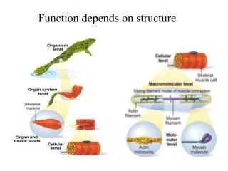
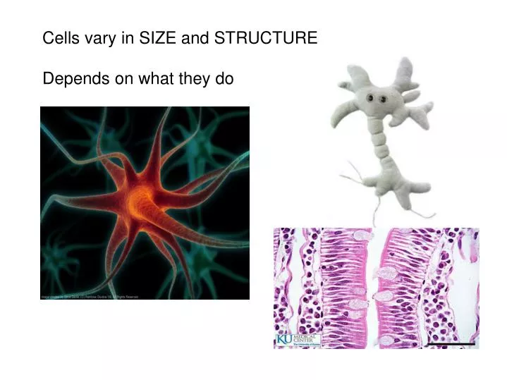
Cells vary in SIZE and STRUCTURE Depends on what they do. All Animal Cells have Nucleus Cytoplasm (cytosol) Cell Membrane. Cell Membrane is selectively permeable. Lipid bilayer (phospholipids) make up the membrane with proteins embedded to help regulate what comes across the membrane.

E N D
Cells vary in SIZE and STRUCTURE Depends on what they do
All Animal Cells have • Nucleus • Cytoplasm (cytosol) • Cell Membrane Cell Membrane is selectively permeable
Lipid bilayer (phospholipids) make up the membrane with proteins embedded to help regulate what comes across the membrane
ORGANELLES- "little organs" within the cell that perform specific functions The nucleus is to the cell what the __________is to a person. The cell membrane is to a cell what the ________ is to a person.
Transport system; canals and channels that connect membrane to nucleus and to organelles within the cell • Smooth ER (lipid synthesis) • Rough ER (contains ribosomes for protein manufacture)
Flattened membranes; function to package and deliver proteins produced by the ribosomes • Proteins are exported in vesicles
Chemical energy from food is converted to a useable form (ATP) • process is called Cellular Respiration • The “powerhouse” of the cell CRISTAE
Lysosomes - contain digestive enzymes to break down substances • Centrosome – forms a spindle during cell division • Vesicles – packaged substances, exported • Microfilaments and Microtubules - cell skeleton (cytoskeleton), maintains shape and functions in movement • Cilia & Flagella
Directs cell activities (the “brain” of the cell) • Contains genetic information (DNA) in the form of chromatin • Also contains a nucleolus – makes ribosomes • Has tiny pores where RNA can exit the nucleus
Diffusion - molecules tend to spread out • Facilitated Diffusion Diffusion Animation
Hypertonic • Hypotonic • Isotonic SALT SUCKS
Active Transport • Exocytosis • Endocytosis (phagocytosis & pinocytosis)
Secretion byexocytosis Proteins in cisterna Rough ER Lysosome fuses withingested substances Cisterna Membrane Transportvesicle Golgi vesicle containingdigestive enzymesbecomes a lysosome Pathway 3 Pathway 2 Golgiapparatus Secretory vesicles Pathway 1 Golgi vesicle containingmembrane componentsfuses with the plasmamembrane Proteins Golgi vesicle containingproteins to be secretedbecomes a secretoryvesicle Plasma membrane Extracellular fluid Figure 3.6, step 12
Extracellularfluid Extracellularfluid Plasmamembrane Cytoplasm Pit Recycling of membraneand receptors (if present)to plasma membrane Transport to plasmamembrane andexocytosis ofvesicle contents Ingestedsubstance Vesicle Lysosome Detachmentof vesicle Release ofcontents tocytoplasm Vesicle containingingested material Plasmamembrane Vesicle fusingwith lysosomefor digestion (a) Active Transport Processes: Endocytosis Figure 3.13a, step 6
Mitosis = nuclear division • Mitosis is followed by cytokinesis (cell division) • The steps of mitosis ensure that each new cell has the exact same number of chromosomes as the original • Interphase = growth phase, • differentiation occurs
Interphase • Prophase • Metaphase • Anaphase • Telophase • IPMAT
The structure of a chromosome Centromere holds two chromatids together
1. chromosomes visible (chromatids) 2. centrioles migrate to the poles 3. nuclear membrane disappears 4. nucleolus disappears 5. spindle forms
1. chromatids separate at the centromere and move to opposite poles
1. chromosomes disappear • chromatin 2. nuclear membrane reforms 3. nucleoli reappears 4. spindle disappears 5. centrioles duplicate
- division of the cytoplasm to form 2 new daughter cells - organelles are divided - daughter cells are genetically identical Cells return to interphase
DIFFERENTIATION occurs as cells multiply and organism develops and grows
Name the phase • Identify X • Identify Y
Name the structure • What is its function?
9. Which beaker(S) contains a solution that is hypertonic relative to the bag A B C D E 10. What will happen to the baggie in the hypertonic solutions?
Nucleus(site of transcription) Cytoplasm(site of translation DNA ) mRNA specifyingone polypeptideis made onDNA template Amino acids mRNA leavesnucleus andattaches toribosome, andtranslationbegins Correct aminoacid attachedto each speciesof tRNA by anenzyme mRNA Nuclear pore Nuclear membrane Synthetaseenzyme Growing polypeptide chain As the ribosomemoves along themRNA, a new aminoacid is added tothe growing proteinchain Met Gly Incoming tRNArecognizes acomplementarymRNA codon callingfor its amino acid bybinding via itsanticodon to thecodon Ser Phe Ala Peptide bond Released tRNAreenters thecytoplasmicpool, ready tobe rechargedwith a newamino acid tRNA “head” bearinganticodon Large ribosomal subunit C G G U U C G C C A U A G U C U A C Portion ofmRNA alreadytranslated Codon Direction of ribosomeadvance; ribosome movesthe mRNA strand alongsequentially as each codonis read Small ribosomalsubunit Protein Synthesis Figure 3.16