Download
1 / 50
500 likes | 848 Views
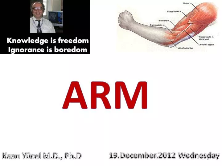
ARM. 19.December.2012 Wednesday. Kaan Yücel M.D., Ph.D. ARM. re gion of the upper limb between the shoulder and the elbow. Superiorly communicates with the axilla . Inferiorly , a number of important structures pass between arm & forearm through cubital fossa.

E N D
ARM • 19.December.2012 Wednesday • Kaan Yücel M.D., Ph.D
ARM regionof the upper limb between the shoulder and the elbow Superiorlycommunicateswiththeaxilla. Inferiorly, a number of importantstructurespassbetweenarm & forearmthroughcubitalfossa.
medial & lateral intermuscular septa Anteriorcompartment – flextheelbowjoint Posteriorcompartment- extendtheelbowjoint ARM Flexion Extension Pronation Supination
Anterior compartment of the arm coracobrachialis, brachialis, and biceps brachii muscles innervated predominantly by musculocutaneous nerve. Posterior compartment triceps brachii muscle innervated by radial nerve.
Coracobrachialis Anelongated muscle in the superomedial part of the arm. Useful landmark for locating other structures in the arm musculocutaneous nerve pierces it distal part of its attachment indicates location of nutrient foramen of the humerus
Coracobrachialis Tip of coracoid process of scapula Middle 1/3 of medial surface of humerus
Coracobrachialis 1. helps flex and adduct the arm 2. stabilize the glenohumeral joint. With deltoid + long head of triceps a shunt muscle, resisting downward dislocation of the head of the humerus, as when carrying a heavy suitcase. Median nerve and/or brachial artery may run deep to coracobrachialis and be compressed by it. Shunt muscle
Coracobrachialis It passes through the axilla and is penetrated and innervated by the musculocutaneous nerve.
shortheadtip of coracoid process of scapulalongheadsupraglenoidtubercle of scapula Biceps brachii Tuberosity of radius and fascia of forearm via bicipitalaponeurosis
Biceps brachii Two heads a single tendon, inserts onto radial tuberosity. and fascia of forearm via bicipitalaponeurosis
Biceps brachii Transverse humeral ligamentconverts the intertubercular grooveinto a canal& holds the tendon of long head of biceps in the groove.
Biceps brachii Triangular membranous band, bicipital aponeurosis, runs from the biceps tendon across the cubital fossa and merges with antebrachial (deep) fascia covering the flexor muscles in the medial side of the forearm. Affords protection for these & other structures in the cubital fossa. Helps lessen the pressure of the biceps tendon on the radial tuberosity during pronation & supination of the forearm.
Biceps brachii • “Three-joint muscle,” crossing & capable of effecting movement at the • Glenohumeral joint • Elbow joint • Radio-ulnar joint primarily acts at the latter two. • Powerful flexor of the forearmat the elbow joint • Most powerful supinator of the forearm when elbow joint is flexed. • Because two heads of biceps brachii muscle cross the glenohumeral joint, the muscle can also flex the glenohumeral joint.
Biceps brachii Elbow flexion approaches 90° and more power is needed against resistance, capable of 2 powerful movements, depending on the position of the forearm. 1) Elbow is flexed close to 90° & forearm supinated: biceps most efficient in producing flexion. 2) Forearm pronated, biceps primary (most powerful) supinator of forearm.
Biceps brachii Innervated by the musculocutaneous nerve. A tap on the tendon of biceps brachii at the elbow is used to testpredominantly spinal cord segment C6.
Brachialis Distal half of anterior surface of humerus Coronoid process and tuberosity ulna Liesbeneath the biceps brachiimuscle Its distal attachment covers the anterior part of the elbow joint.
Brachialis Main flexor of the forearm The only pure flexor, producing the greatest amount of flexion force primarilyresponsible for sustaining the flexedposition workhorseof the elbowflexors
Brachialis Innervation predominantly by musculocutaneous nerve. A small component of the lateral part is innervated by the radial nerve.
Triceps brachii The only muscle of the posterior compartment • longhead • infraglenoidtubercle of scapula • medial head & lateralheadsposterior surface of humerus, superior to radial groove • Proximal end of olecranon of ulna and fascia of forearm
Triceps brachii Because its long head crosses the glenohumeral joint, the triceps helps stabilize the adducted glenohumeral joint by serving as a shunt muscle, resisting inferior displacement of the head of the humerus. The long head also aids in extension and adduction of the arm, but it is actually the least active head. Medial head : workhorse of forearm extension, Lateral head : strongest but is recruited into activity primarily against resistance.
Triceps brachii Innervation of by branches of the radial nerve. A tap on the tendon of triceps brachii tests predominantly spinal cord segment C7.
Brachial artery The major artery of the arm Found in the anterior compartment Continuation of axillary artery at the lower border of teres major Terminates distal to the elbow joint, opposite to neck of radius dividing into radial & ulnar arteries.
Brachial artery Relatively superficial and palpable throughout its course. Lies anterior to triceps & brachialis. As it passes inferolaterally, accompanies the median nerve.
Brachial artery Proximal arm lies on the medial side. Distal arm, it moves laterally. Named Branches Superior ulnar collateral artery Inferior ulnar collateral artery contribute to a network of arteries around the elbow joint. Profunda brachii artery Nutrient arteries to the humerus
Brachial artery Deep artery of the arm (L. arteria profunda brachii) Largest branch & most superior origin Accompanies radial nerve along the radial groove Terminates by dividing into middle & radial collateralarteries
2 main superficial veins of the arm cephalicand basilic veins. Cephalicvein – lateralside intoaxillaryvein Basilicvein- medialside Basilicvein+ Brachialveins Axillaryvein
NERVES IN THE ARM
4 main nerves pass through the arm: • Median • Ulnar • Musculocutaneous • Radial
Musculocutaneous nerve Leaves the axilla and enters the arm by passing through the coracobrachialis muscle. Passes diagonally down the arm between biceps brachii & brachialis.
Musculocutaneous nerve • Throughcoracobrachialis • Diagonallydown the arm • in the plane between biceps brachii&brachialis • Emergeslaterally totendonof bicepsbrachii@ theelbow • lateral cutaneous nerve of forearm • motor innervation to all muscles @ anterior compartment of the arm; • sensory innervation to skin @ lateral surface of theforearm
Median nerve Enters the arm from axilla @ inferior margin of teres major muscle. Passes vertically down the medial side of arm in the anterior compartment Related to brachial artery throughout its course: No major branches in the arm, or in the axilla.
Ulnar nerve Enters the arm with the median nerve and axillary artery. Passes distally from the axilla anterior to the insertion of the teres major and to the long head of the triceps, on the medial side of the brachial artery. In the middle of the arm, penetrates the medial intermuscular septum and enters the posterior compartment. Passes into the anterior compartment of the forearm.
Ulnar nerve Posterior to the medial epicondyle, where the ulnar nerve is referred to in lay terms as the “funny bone,” it is superficial, easily palpable, and vulnerable to injury. .
Radial nerve Supplies all the muscles in the posterior compartment of the arm (and forearm). Enters the arm by crossing the inferior margin of teres major muscle. Entersthe posterior compartment of the armthroughtriangularinterval. profundabrachiiartery
Radial nerve Muscular and cutaneous branches in the arm Muscular branches include those to Triceps brachii Brachioradialis Extensor carpi radialis longus muscles. Contributes to innervation of lateral part of brachialis muscle.
Radial nerve Cutaneous branches Inferior lateral cutaneous nerve of arm skin over lateral & anterior aspects of the lower part of the arm. Posterior cutaneous nerve of forearm penetrates through the lateral head of triceps brachii muscle & overlying deep fascia to become subcutaneous.
Radial nerve Anterior to lateral epicondyle, divides into Deep branch (muscular & articular) Superficial branch (cutaneous- dorsum of the hands & fingers)