Download

1 / 49
590 likes | 1.26k Views
The Excitation-contraction coupling in skeletal muscle. Department of Animal Science & Technology National Taiwan University De-Shien Jong. Outline. Excitation-contraction coupling of skeletal muscle Charge movement & Calcium release Poor man ’ s fura-2 Model of Ca release

E N D
The Excitation-contraction coupling in skeletal muscle Department of Animal Science & Technology National Taiwan University De-Shien Jong
Outline • Excitation-contraction coupling of skeletal muscle • Charge movement & Calcium release • Poor man’s fura-2 • Model of Ca release • Effects of Ca on kinetics of charge movement • Model of charge movement
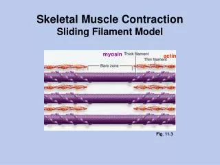
Schematic drawing of skeletal muscle Peachey, 1965
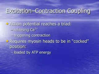
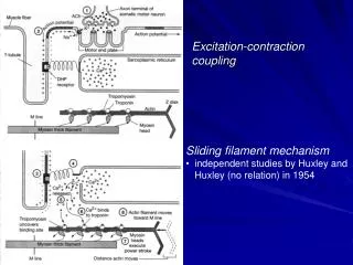
Excitation-Contraction coupling • Skeletal muscle • Action potential → Motor neuron → Neuromuscular junction → muscle surface → Transverse tubular system (T-system) → Triad → Dihydropyridine Receptors (DHPR) → Ryanodine Receptors (RyR) → Ca2+ release from SR → bind to troponin → induce muscle contraction • How does DHPR and RyR talk to each other?
Excitation-Contraction coupling • Cardiac muscle • Action potential → Triad (or Diad) → DHPR → Ca2+ entry → Ca2+ bind to RyR → Ca2+ induce Ca2+ Release → Ca2+ bind to contractile protein → muscle contraction • DHPR is L-type voltage-gated Ca2+ channel which has two isoforms : Skeletal type & Cardiac type
Ryanodine Receptor • RyR has two isoforms in amphibian muscle : a & b • RyR has three isoforms in mammalian muscle : RyR1, RyR2, and RyR3 • RyR1 mainly in skeletal muscle • RyR2 in cardiac muscle • RyR3 in most other cells (i.e. brain)
Ferguson et al. 1984 Felder & Franzini-Armstrong, 2002
Charge movement & Calcium release Dr. W. Knox Chandler Dr. Paul C. Pape Dr. Steve M. Baylor
Chandler et al. 1976 Miledi et al. 1977, Caputo et al. 1984
Effects of increased [Ca2+]i • Bind to contractile protein troponin • Bind to various intrinsic Ca buffers • Activate additional release of Ca from the SR (Ca induced Ca release) • Reduce additional Ca release from the SR (Ca inactivation of Ca release) • Bind to Ca indicators
Experimental methods • A cut frog muscle fiber was mounted on a double Vaseline-gap chamber • Extracellular and intracellular solutions contain ion replacement to eliminate ionic currents • Internal solution contained 20 mM EGTA plus 1.76 mM Ca2+, which expecting to catch all the Ca released from the SR
480 nm : isobestic wavelenth of phenol red 570 nm : a wavelength at which phenol red is sensitive to pH 690 nm : a wavelength not absorbed by phenol red Fractional phenol red in the nonprotonated form f f = (r - rmin) / (rmax - rmin) where r = Aind(570) / Aind(480) pH = pk + log (f / (1 – f))
Advantages of EGTA-phenol red method • The EGTA-phenol red method estimates SR Ca release reliably (~ 96%) and Rapidly (<0.1 ms). • The change in pH does not alter physiological condition and the buffering power b is stable. • The dissociatin rate of Ca and EGTA, k-1, is small (~ 1 s-1) compared to that of Ca and fura-2 (~10-30 s-1).
k1[EGTA]-1 = 22 ms So the D[Ca] signal estimated from DpH would be the same amplitude as the measurement with PDAA
D[Ca] = f / (4 p DCa r) * exp ( -r / lCa) Where lCa = { DCa / k1[EGTA]R}1/2 = 81 nm
Time course of D[Ca] after a step change in f from a single point source in an isotropic infinite medium.
Case 1 Case 2 an = f n a0, bn = f n b0 an = f n a0, bn = f -n b0
Case 3 Exp. Data an = f’f n a0, bn = f’f -n b0