Download
1 / 64
680 likes | 882 Views
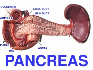
Imaging Approach to Pancreas tumours. Ryno vd Berg Dept Radiology Kimberley hospital 13/4/2012. Retroperiteal organ, except for tail in splenorenal lig at L1 level Head(uncinate process), neck, body & tail.

E N D
Imaging Approach to Pancreas tumours Rynovd Berg Dept Radiology Kimberley hospital 13/4/2012
Retroperiteal organ, except for tail in splenorenal lig at L1 level • Head(uncinate process), neck, body & tail. • Size decreases with age; rough guide: Head≤3.5cm & Rest ≤2.5cm. Pancreatic duct 1-3mm. • Bloodsupply from splenic, R gastro-omental & Sup mesenteric aa. • Drainage to portal system. Endocrine part (2%): Langerhans islets: α-,β-,δ- & PP-cells Islets clustered together crossed by network of capillaries Exocrine part: Acinar cells producing & secreting digestive enzymes Alkaline juice secreted by ductal cells Secreted into duodenum for digestion with bile Pancreas:
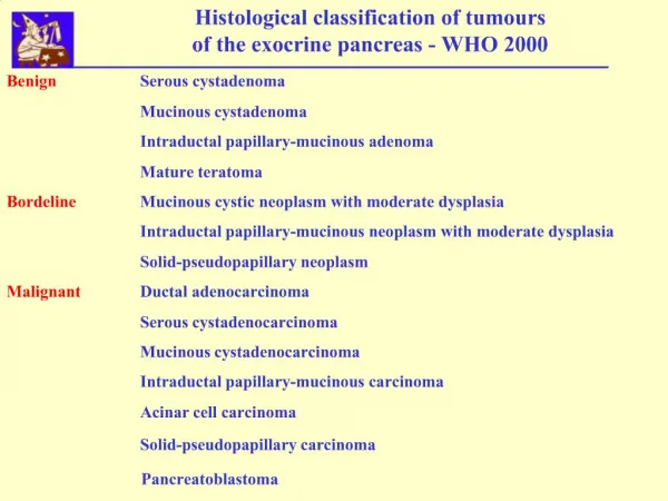
Solid lesions Malignant Benign • Cystic lesions Unilocular simple Small cystic Septated • Solid with cystic components Imaging classification:
Malignant: 1.AdenoCA* 2.Neuroendocrine* 3.Solid Pseudopapillary* 4.Panceaticoblastoma 5.Lymphoma 6.Mets to Pancreas* 7.Very rare: Epthelial: Acinar cell, Giant cell & Colloid CA Mesenchymal: Granular cell CA, Fibrous histiocytoma, Juvenile hemangioendothelioma, Fibroma, Inflam Myoblastic tumor & Sarcoma* Mixed: Squamous cell & Mixed endo- exocrine tumors Benign: 1.Focal Pancreatitis 2.Fatty infiltration/replacement 3.Intrapancreatic accessory spleen 4.Pancreas lobulation/Cong anomalies 5.Miscellaneous: Pancreatic Sarcoidosis Castleman’s Disease Solid Pancreas lesions: *May have cystic degeneration
85-95% of all Pancreatic malignancies • Age 60-80y, Males(2:1) • Head – 60-70%, Body – 10-20%, Tail- 5-10% • Poor prognosis (5-y survival < 5%) • Non-resectable in 75% on presentation • Surgery only cure (5-y survival 20%) • Need appropriate pt selection to prevent unnecessary surgery Pancreatic Adenocarcinoma
CT: Hypovascular with low attenuation best seen in arterial phase Porto-venous phase to detect mets & evaluate surrounding veins 10% Isodense, look for secondary signs (Abn pancreas shape, ductal obstruction, vascular invasion) Mets commonly to liver, lymphnodes, peritoneum & lung Double duct sign Isodense mass in head causing abrupt distal narrowing & dilatation of CBD
MRI: Fibrotic nature – Low on T1 & T2 Often thin peritumour ring of increased enhancement Superior to CT for detecting small tumours & Mets Accuracy for detecting & staging of AdenoCA 90-100%
Endoscopic US: Role in detecting small tumours (2-3mm) and help clarify equivocal CT & MR findings Ill-defined, heterogenous hypoechoic mass 98% sensitivity in detecting adenoCA However operator dependent & narrow field of view for assessing local invasion PET: Increased uptake & retention of FDG Sensitivity 85-100%, Specificity 84-93% Biggest impact ability to detect small metastases False negatives: Mucinous tumours, Necrotic tumours, Peritoneal mets < 1cm & hyperglyceamia False positives: Inflammatory tissue
Criteria for defining the respectability of Pancreatic AdenoCA Resectible: 1. No mets 2. No abutment, distortion, tumour thrombus or encasement of SMV/PV 3. Clear fat planes around CA, SMA & HA Borderline resectable: 1. Abutment or encasement of SMV/PV (No impingement/ narrowing of lumen) 2. Short segment occlusion from tumour thrombus/ encasement, but allowing safe resection & reconstruction 3. Encasement of Gastroduodenal a up to HA (only short segment encasement/ abutment of HA & no involvement of CA) 4. <180° Abutment of SMA circumference • Not resectable: • Mets • Lymphnodemets beyond the field of resection • >180° Encasement of SMA • Unreconstructable occlusion of SMV/PV • Aortic invasion/ encasement National Comprehensive Cancer Network- Radiographics, July 2011
Previously Islet cell tumours 1-5% of all Pancreas tumours, Age 51-57y, Equal both genders Mostly sporadic, but may be associated with MEN type 1, Von Hippel-Lindau, Neurofibromatosis type 1 & Tuberous Sclerosis Classified into Functioning & Non-functioning Further histologically: • Well differentiated benign features • Well differentiated uncertain • Well differentiated carcinoma • Poorly differentiated carcinoma Pancreatic Neuroendocrine tumours
Insulinoma: • Most common NET (60-75%) • 10% Malignant • M:F = 2:3 Age: 40-60 yr • Associated with MEN I in 10% • No predilection for any part of the pancreas • Mostly <2cm • Classically hypervascular • Surgical resection is curative • Localization of tumour prior to surgery: • Endoscopic US / intra-operative US • Angiography • Intra-Arterial Calcium Stimulation with Hepatic Venous Sampling • Catheterize • Superior mesenteric artery (SMA) • Gastroduodenal artery (GDA) • Splenic artery (SpA) • Twofold rise in insulin levels following calcium gluconate injection in SMA or GDA = tumour is located in the pancreatic head or uncinate process • Twofold rise after injection into the SpA suggests caudal location
Gastrinoma: • 2nd most common NET • 80% in patients < 20yr M>F • Associated with MEN I in 50-60% • 50-60% malignant • Tumour size does not appear to be associated with biological behaviour or clinical course • Lymph node or liver metasteses present in 70-80% of cases at diagnosis • Zollinger –Ellison syndrome: Hypersecretion of gastrin leading to Acid secretion with peptic ulceration & diarrhoea • Average tumour size 3,5cm (up to 15cm) • Tend to be less vascular than insulinoma • More often multiple compared to insulinoma • Associated metasteses • Associated gastric wall thickening, indicating peptic ulcer disease and gastric hyperplasia due to effects of gastrin can be helpful in diagnosis
Non-Functioning NET: • 3rd most common • Histological from alpha or beta cells • Mean age 57 yrs • Symptoms due to mass effect (present like adenocarcinoma) • Predominantly in pancreatic head • 90% malignant at presentation • Imaging • Large in size 3- 25cm • Large, well-defined masses with moderate to strong enhancement • No invasion of adjacent vessel • Associated with cystic degeneration and calcifications • More easily detected by mass effect compared to functioning tumors
Glucagonoma: • Uncommon tumour • Middle age M<F • Associated with MEN • Secrete excessive amounts of glucagon and cause a syndrome characterized by dermatitis, stomatitis, weight loss, and anaemia(fatigue) • ( 4D syndrome = dermatosis, diarrhoea, depression, dvt ) • Increased glucagon leads to glucose intolerance (DM) and cachexia (catabolic effects) • Third of patients with glucagonoma syndrome have secondary thromboembolic phenomena • This feature is unique among the different NET • Predominantly in pancreas body & tail • Large tumour (>5cm in 70%) with solid and necrotic elements • Hypervascular in 90 % • 60-80% malignant transformation • 60% liver metasteses at presentation
VIPoma: • Excretes Vasoactive Intestinal Peptide • Acting directly on cyclic AMP within epithelial cells in bowel • Relaxes vascular smooth muscle + causes electrolyte secretion • WDDH syndrome: • Watery diarrhoea + hypokalaemia + hypochlorhydria • “pancreatic cholera” / Verner-Morrison syndrome • >50% malignant transformation
Somatostatinoma: • May be associated with NF1 • symptoms reflect the general inhibitory action of somatostatin on global gastroenteropancreatic function • often have history findings consistent with diabetes mellitus, which is probably secondary to the inhibitory action of somatostatin on insulin and glucagon release • Inhibition of the action of cholecystokinin by somatostatin causes relative biliary stasis and the formation of gallbladder calculi • diarrhea and/or steatorrhea, both of which are likely to be caused by the inhibition of pancreatic enzyme and bicarbonate secretion • Predominantly in • Pancreas head • Duodenum at ampulla of Vater • Tumour size 0,6-20cm ( avg >4cm ) • >50% chance of malignant transformation • 60% liver/lymph node metasteses at presentation
CT: Most distinctive feature: rich blood supply – enhancing more rapid + intense than surrounding tissue in arterial phase Homogenous enhancement of tumours < 2cm & more heterogenous for larger tumours which can be ring-like Porto-venous phase show no typical pattern Metastases often follow same enhancement pattern as primary Sensitivity 94% MR: Generally have longer T1&T2 relaxation than other tumours and normal pancreas Low on T1 and intermediate to high on T2 Sensitivity 85-94% Imaging approach to NET
Endoscopic US: Sensitivity 94%, but combined with CT up to 100% Also useful in detecting duodenal gastrinomas & used in u/s guided FNA SPECT/CT: Radiolabeled Octreotide (Somatostatin analog) taken up by most NET, except insulinomas Sensitivity 90% Specificity 80%, however helpful in detecting mets PET: Role in detecting poorly differentiated NET, which have a high proliferative rate (High FDG uptake), compared to well differentiated NET with low proliferation rates Differentiating NET from AdenoCA:
Insulinoma: Solid mass with hypervascular periphery Splenic arteriogram showing tumour in pancreatic tail
Gastrinoma: heterogeneous mass in the head of the pancreas with cystic degeneration and shell-like dense calcification. (b) Wall of the gastric antrum is hypertrophied
Solid cystic papillary epithelial-, Papillary cystic-, Solid and cystic-/ Franz tumour 1-2% of Pancreas tumours Age range 10-74y (mostly young adults), female predominance (9:1), African & Asian Low malignant potential with excellent prognosis post resection Mets uncommon (7-9%): Liver, omentum, peritoneum Typically large (mean 9cm), slow-growing, well encapsulated mass Common in pancreas tail & tend to displace rather than invade Solid Pseudopapillary tumour
CT: Hypodense pseudocapsule (compressed pancreas tissue & fibrosis) Peripheral heterogenous enhancement during arterial phase followed by progressive non-uniform enhancement (generally less than surrounding pancreas) 10-20% fluid-fluid or fluid-debris level 30% Peripheral calcifications MR: Capsule low T1&T2 Internal haemorrhage & cystic degeneration due to fragile vascular network of tumour Subacute bleeding (high on T1), Chronic bleed (Low on T1+T2)
Main differential to consider: Well encapsulated solid tumour, low on T1 & interm-high on T2
0.2% of all pancreas tumours, but the most common in children (mean age 5y) Male predominance (1.3:2.7), Asian (>50% of all cases) Raised α-FP in 25-33% Slow growing & usually manifest as asymptomatic large mass (mean 10cm) Adrenocorticotropic hormone secretion have been documented (Cushing’s / Inapp ADH) Due to large size often not possible to identify organ of origin and generally require biopsy to diagnose Rarely cause bile/ duodenal obstruction due to soft gelatinous consistency Metastases: Liver, lymphnodes, lung, bone, mediastinum, peritoneum & omentum US: Heterogeneous mass with hypoechoic cystic spaces (necrosis) and hyperechoic internal septae Occasionally only hypoechoic solid mass CT: Generally multiloculated heterogeneous mass with enhancing septae Calcifications if present rimlike or clustered Mild contrast enhancement MR: Low-interm T1 & High T2 Pancreatoblastoma
Pancreatoblastoma in 5y old: US: Large heterogenous mass with cystic spaces encasing CA & displacing the Portal vein CT: Solid mass with cystic components displacing the Portal confluence & stomach anteriorly
Most commonly B-cell type of Non-Hodgkin lymphoma Primary: <2% of Extranodal lymphomas & 0.5% of Pancreatic tumours Secondary: In 30% of widespread NH Lymphomas Age 35-75y, Immunocompramised Two morphological patterns: 1.Focal well circimscribed: 80% in head Mean size 8cm CT: Uniform hypodense MR: Low T1, Interm T2 Faint contrast enhancement 2.Diffuse form: CT: Infiltrative with enlargement & poorly defined of pancreas MR: Low T1 + T2 Homogeneous contrast enhancement Important to distinguish from AdenoCa, because better prognosis & Chemo as 1st line treatment: • Mild pancreatic duct dilatation & CBD dilatation more common • Enlarged lymphnodes below the level of the renal vein • Invasive – infiltrates retroperitoneum, surrounding organs & GIT • Vascular invasion less common • Calcifications & necrosis not feature Pancreatic Lymphoma
Primary Pancreas Lymphoma: Pt 10y post renal transplant Focal hypodense tumour in head Secondary Pancreas Lymphoma: Local invasion of pancreas tail from lymphomatous infiltration of the spleen Also extensive retroperitoneal lymphadenopathy
2-5% of Pancreatic neoplasms Mostly from: Renal cell CA, Lung CA, Breast CA, Colorectal CA & Melanoma US: Hyper- / hypoechoic CT: Hypo-/Isodense MR: Low T1 & high on T2 Follow enhancement pattern of primary >1.5cm mostly peripheral enhancement with central necrotic area & smaller lesions mostly homogeneous enhancement Renal cell CA hypervascular, need to differentiate from NET Rest mostly hypovascular, need to diff from AdenCA Past medical history very important Metastases to the Pancreas
Mets from Renal cell CA Asymptomatic pt had nephrectomy 20y ago Hypervascular tumour with cystic-necrotic degeneration & intratumour vessels
Chronic pancreatitis: Focal inflammatory mass, often in head Account for 5-10% of pancreatectomies for presumed malignancy Difficault to distinguish from AdenoCA, even histologically Focal Pancreatitis
Focal chronic pancreatitis Focally enlarged head with irregular contour & internal calcifications Remained stable over 3y after follow up imaging
Autoimmune Pancreatitis: (AIP) 25% of focal pancreatits May occur alone or with other autoimmune disorders; most patients have increased IgG and antinuclear antibody levels US: focal hypoechoic, diffuse enlargement (“sausage”)/ normal CT: diffuse enlargement , loss of normal surface indentations, tail retracted from splenichilum, capsule-like rim enhancement, peripancreaticadenopathy, no calcification or vascular encasement MRCP/ERCP: diffuse irregular narrowing of pancreatic duct(often >3cm long), CBD stricture Diffusely enlarged pancreas with hypodense capsule-like rim
Groove pancreatitis: Uncommon form of focal pancreatitis involving pancreaticoduodenal groove Two forms: 1. Segmental – Involves head with scar tissue in the groove 2. Pure – Affects the groove, but spares the head May manifest as duodenal or biliary obstruction CT: Sheetlike hypodense fibrotic scar tissue in groove, delayed contrast enhancement MR: Low on T1, Interm-high on T2 Associated with smooth stricture of pancreatic part of CBD & Wall thickening + cystic dysplasia of duodenum
Common finding in elderly & obese, but usually diffuse Also associated with chronic pancreatitis & Cystic fibrosis Has predilection for anterior part of head, sparing posterior part & uncinate process US: Fat – Hyperechoic & spared areas hypoechoic CT: If macroscopic will show negative HU on uncontrasted scan (contrast spread between fat increasing attenuation) Absence of mass effect MR: Modality of choice Macroscopic fat easily detected on fat-sat sequences Microscopic fat detected with chemical shift imaging Dual phase T1 weighted GRE series: Areas show high on the in-phase & signal loss on opposed-phase sequence Hypoechoic area of fatty sparing in hyperechoic fatty pancreas Fatty infiltration-replacement
Failed fusion of splenic anlage in dorsal mesogastrium Typically 1-3cm, well-defined ovoid mass in tail US: Homogeneous, mildly echogenic. May show posterior acoustic enhancement Vascular hilum sometimes seen with doppler CT: Greater enhancement similar to spleen with same tigroid pattern MR: Lower on T1, Higher on T2 Need to diff from other vascular lesions: Superparamagnetic iron-oxide MR: Phagocytosed by reticuloendothelial cells, leading to decreased signal on post-contrast series Scintography: Splenic tissue traps 90% 99m-Tc Heat damaged RBC Intrapancreaticaccesory spleen
Well-defined oval mass with same intensity as spleen Same enhancement pattern SPECT/CT with 99m-TC HDRBC
Pancreatic lobulation: Bifid pancreas tail: Tuber omentale: Focal prominence of anterior pancreatic surface left of sup mesenteric vessels Congenital anomalies
Sarcoidosis: Idiopathic systemic granulomatous disorder Pancreatic involvement very rare, only 19 biopsy-proven cases in literature Solitary/ multiple masses, may reach 6-7cm Peripancreaticlymphnodeenlargent US: Hypoechoic CT: Hypodense & non-enhancing MR: Low T1, High T2 & Hypoenhancing History & other findings Castleman Disease: Rare angiofollicularlymphnode hyperplasia Only 10 cases of pancreatic involvement documented CT: Solid, well-encapsulated mass with strong enhancement (may be ringlike) May have calcifications & cystic changes Very rare solid masses
Benign: Serous cystadenoma Cystic teratoma Inflammatory: Pseudocyst, Abscess, Echinococcus True epithelial cysts: Von Hippel-Lindau Adult PCKD Cystic fibrosis Rare: Lymphangioma Hemangioma Paraganglioma Malignant/ Potentially malignant: Mucinous cystic neoplasm Intraductal papillary mucinous neoplasm (IPMN) Solid with cystic degeneration: AdenoCa Solid pseudopapillary tumour Neuroendocrine tumours Mets Sarcoma Cystic teratoma Cystic pancreatic lesions
Pseudocyst, IPMN, Serous cystadenoma, Epithelial cysts Serous Cystadenoma Mucinous cystic neoplasm IPMN AdenoCa Solid pseudopapillary tumour Neuroendocrine tumours Mets Sarcoma Cystic teratoma Mucinous cyst adenoma, IPMN Morphological classification
Pseudocyst: No epithelial lining, but fibrous wall surrounding pancreatic fluid, cellular debris & blood Most common cystic mass Complication of acute or chronic pancreatitis or pancreatic trauma US: Usually well defined, smooth-walled anechoic cyst May be multilocular with internal septations with internal echoes or fluid-fluid levels if heamorrhage or infected CT: Usually well defined round thin/thick walled fluid collection with mild rim enhancement Inflammatory cysts
Pancreas abscess Haemorrhagic pseudocyst
Echinococcus cyst: Rare, liver & lung involvement much more common Uni-/multilocular Peripheral enhancement Peripheral calcifications my be present
Ussually multiple small simple cysts PCKD with cyst in pancreas Von Hippel-Lindau disease True epithelial cysts