Download
1 / 1
10 likes | 162 Views
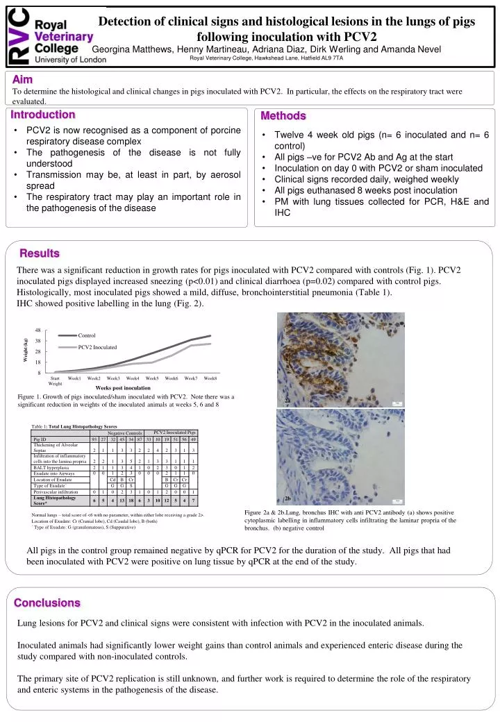
Detection of clinical signs and histological lesions in the lungs of pigs following inoculation with PCV2 . Georgina Matthews, Henny Martineau, Adriana Diaz, Dirk Werling and Amanda Nevel Royal Veterinary College, Hawkshead Lane, Hatfield AL9 7TA .

E N D
Detection of clinical signs and histological lesions in the lungs of pigs following inoculation with PCV2 Georgina Matthews, Henny Martineau, Adriana Diaz, Dirk Werling and Amanda Nevel Royal Veterinary College, Hawkshead Lane, Hatfield AL9 7TA To determine the histological and clinical changes in pigs inoculated with PCV2. In particular, the effects on the respiratory tract were evaluated. Aim Introduction Methods • PCV2 is now recognised as a component of porcine respiratory disease complex • The pathogenesis of the disease is not fully understood • Transmission may be, at least in part, by aerosol spread • The respiratory tract may play an important role in the pathogenesis of the disease • Twelve 4 week old pigs (n= 6 inoculated and n= 6 control) • All pigs –ve for PCV2 Ab and Ag at the start • Inoculation on day 0 with PCV2 or sham inoculated • Clinical signs recorded daily, weighed weekly • All pigs euthanased 8 weeks post inoculation • PM with lung tissues collected for PCR, H&E and IHC There was a significant reduction in growth rates for pigs inoculated with PCV2 compared with controls (Fig. 1). PCV2 inoculated pigs displayed increased sneezing (p<0.01) and clinical diarrhoea (p=0.02) compared with control pigs. Histologically, most inoculated pigs showed a mild, diffuse, bronchointerstitial pneumonia (Table 1). IHC showed positive labelling in the lung (Fig. 2). Results Figure 1. Growth of pigs inoculated/sham inoculated with PCV2. Note there was a significant reduction in weights of the inoculated animals at weeks 5, 6 and 8 2a 2b Figure 2a & 2b.Lung, bronchus IHC with anti PCV2 antibody (a) shows positive cytoplasmic labelling in inflammatory cells infiltrating the laminar propria of the bronchus. (b) negative control All pigs in the control group remained negative by qPCR for PCV2 for the duration of the study. All pigs that had been inoculated with PCV2 were positive on lung tissue by qPCR at the end of the study. Lung lesions for PCV2 and clinical signs were consistent with infection with PCV2 in the inoculated animals. Inoculated animals had significantly lower weight gains than control animals and experienced enteric disease during the study compared with non-inoculated controls. The primary site of PCV2 replication is still unknown, and further work is required to determine the role of the respiratory and enteric systems in the pathogenesis of the disease. Conclusions