Download
1 / 23
230 likes | 376 Views
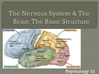
Structure of the Nervous System. The Central nervous System (CNS) Parts of the nervous system that are encased in bone Brain Spinal Cord. The Peripheral nervous System (PNS) All the spinal nerves that innervate the skin, joints, muscles , etc. and under voluntary control : Somatic PNS

E N D
The Central nervous System (CNS) • Parts of the nervous system that are encased in bone • Brain • Spinal Cord
The Peripheral nervous System (PNS) All the spinal nerves that innervate the skin, joints, muscles, etc. and under voluntary control: Somatic PNS Neurons thatinnervate internal organs, blood vessels, glands, etc. and are involuntary: Visceral PNS or Autonomic Nervous System (ANS)
Anatomical Reference Dorsal Caudal/ Posterior Rostral/ Anterior Medial Lateral Ventral Ipsilateral & Contralateral
Anatomical Reference Horizontal Section Midsagittal Section Coronal/Transverse Section
Side (Lateral) view Cerebrum Cerebellum Brain Stem Spinal Cord Top (Dorsal) view Sagittal fissure Cerebral hemispheres What would a Midsagittal view be?
Dura mater (Hard Mother) Subdural space Arachnoid membrane Subarachnoid space Pia mater (Gentle Mother) Artery Brain Ventricles CSF (Cerebro-spinal Fluid)
Early development of nervous system in embryo Neural Plate Neural Groove Neural Tube Fuse Dorsally Rostral Prosencephalon (forebrain) Mesencephalon (midbrain) Rhombencephalon (hindbrain) Caudal
Development of nervous system in embryo Telencephalon (2 cerebral hemespheres) Diencephalon (between brain) Mesencephalon (Midbain)
Development of nervous system in embryo Telencephalon Diencephalon Corpus Callosum Cerebral Cortex Thalamus Hypothalamus Coronal Section Lateral Ventricles & Third Ventricle
Development of nervous system in embryo Midbrain: becomes Tectum (roof) Tegmentum (floor) Hindbrain: becomes Cerebellum Pons Medulla
Virtual Hospital: University of Iowa health Care http://www.vh.org/adult/provider/anatomy/BrainAnatomy/BrainAnatomy.html
Cerebellum 1. Flocculus 2. Uvula of vermis 3. Tonsil 4. Biventral lobule 5. Pyramis of vermis 6. Tuber of vermis 7. Inferior semilunar lobule
Brain Stem & Cerebellum 1. Oculomotor nerve 2. Interpeduncular fossa 3. Basis pedunculi 4. Basilar sulcus of pons 5. Motor (minor) root of trigeminal nerve 6. Sensory (major) root of trigeminal nerve 7. Abducens nerve 8. Middle cerebellar peduncle 9. Vestibulocochlear nerve 10. Facial nerve 11. Flocculus 12. Choroid plexus protruding through lateral aperture of 4th ventricle (foramen of Luschka) 13. Glossopharyngeal nerve 14. Vagus nerve 15. Accessory nerve 16. Olivary nucleus 17. Pyramidal tract 18. Hypoglossal nucleus 19. Pyramidal decussation
Cerebral hemisphere Dorsal View 1. Frontal pole 2. Superior frontal sulcus 3. Middle frontal gyrus 4. Superior frontal gyrus 5. Precentral sulcus 6. Longitudinal cerebral fissure 7. Precentral gyrus 8. Postcentral gyrus 9. Central sulcus 10. Postcentral sulcus 11. Occipital pole
Cerebral hemisphere Ventral View 1. Frontal pole of left cerebral hemisphere 2. Olfactory bulb 3. Olfactory tract 4. Orbital gyri and sulci 5. Straight gyrus 6. Temporal pole of left cerebral hemisphere 7. Olfactory trigone 8. Optic nerve 9. Optic chiasma 10. Anterior (rostral) perforated substance 11. Optic tract 12. Tuber cinereum with infundibulum 13. Oculomotor nerve 14. Mamillary body 15. Uncus of parahippocampal gyrus 16. Basis pedunculi 17. Basilar sulcus of pons 18. Trigeminal nerve 19. Abducens nerve 20. Pyramid of medulla oblongata 21. Facial nerve 22. Vestibulocochlear nerve 23. Glossopharyngeal nerve 24. Vagus nerve 25. Cranial roots of accessory nerve 26. Spinal roots of accessory nerve 27. Rootlets of hypoglossal nerve 28. Flocculus 29. Ventral rootlets of 1st cervical spinal nerve 30. Pyramidal decussation
Cerebral hemisphere Lateral View 1. Superior frontal gyrus 2. Superior frontal sulcus 3. Central sulcus 4. Precentral gyrus 5. Postcentral gyrus 6. Supramarginal gyrus 7. Angular gyrus 8. Postcentral sulcus 9. Parieto-occipital sulcus 10. Superior parietal lobule 11. Intraparietal sulcus 12. Precentral sulcus 13. Middle frontal gyrus 14. Inferior frontal sulcus 15. Inferior frontal gyrus 16. Anterior ascending ramus of lateral sulcus 17. Transverse temporal gyrus 18. Anterior horizontal ramus of lateral sulcus 19. Superior temporal gyrus 20. Superior temporal sulcus 21. Middle temporal gyrus 22. Stem of lateral sulcus 23. Inferior temporal sulcus 24. Inferior temporal gyrus 25. Preoccipital notch 26. Posterior branch of lateral sulcus 27. Triangular part of inferior frontal gyrus 28. Opercular part of inferior frontal gyrus
Cerebral hemisphere Midsagittal View 1. Medial frontal gyrus 2. Cingulate sulcus 3. Cingulate gyrus 4. Central sulcus 5. Paracentral lobule 6. Callosal sulcus 7. Isthmus of cingulate gyrus 8. Subparietal sulcus 9. Precuneus 10. Parieto-occipital sulcus 11. Cuneus 12. Calcarine sulcus or fissure 13. Rostrum of corpus callosum 14. Genu of corpus callosum 15. Trunk of corpus callos 16. Splenium of corpus callosum 17. Choroid plexus in interventricular foramen 18. Interthalamic adhesion 19. Habenular trigone 20. Hypothalamic sulcus 21. Pineal body 22. Anterior (rostral) commissure 23. Tectum of midbrain 24. Mamillary body 25. Medial longitudinal fasciculus 26. Choroid plexus of 4th ventricle
Cerebral hemisphere Midsagittal View 1. Medial frontal gyrus 2. Cingulate gyrus 3. Central sulcus 4. Paracentral lobule 5. Cingulate sulcus 6. Callosal sulcus 7. Subparietal sulcus 8. Precuneus 9. Parieto-occipital sulcus 10. Cuneus 11. Isthmus of cingulate gyrus 12. Lingual gyrus 13. Calcarine sulcus or fissure 14. Medial occipitotemporal gyrus 15. Collateral sulcus 16. Parahippocampal gyrus 17. Uncus of parahippocampal gyrus 18. Rhinal sulcus 19. Subcallosal area 20. Paraterminal gyrus 21. Indusium griseum 22. Rostrum of corpus callosum 23. Genu of corpus callosum 24. Trunk of corpus callosum 25. Splenium of corpus callosum 26. Fimbria of hippocampus 27. Cut surface of thalamus 28. Anterior (rostral) commissure 29. Interthalamic adhesion 30. Column of fornix 31. Septum pellucidum
Cerebral hemisphere Midsagittal View 1. Corona radiata 2. Head of caudate nucleus 3. Body of caudate nucleus 4. Tail of caudate nucleus 5. Anterior thalamic peduncle 6. Stria terminalis 7. Anterior nuclear group of thalamus 8. Dorsal lateral thalamic nucleus 9. Stria medullaris thalami 10. Habenular nucleus 11. Pulvinar 12. Mamillothalamic fasciculus 13. Anterior (rostral) commissure 14. Column of fornix 15. Hypothalamic nuclei 16. Substantia nigra 17. Red nucleus 18. Habenulo-interpeduncular tract 19. Temporal pole 20. Optic tract 21. Mamillary body 22. Interpeduncular nucleus 23. Medial lemniscus 24. Median section of pons 25. Lower lip of parieto-occipital sulcus 26. Cuneus 27. Calcarine sulcus
Cerebral hemisphere Coronal View 1. Body of corpus callosum 2. Frontal horn of lateral ventricle 3. Septum pellucidum 4. Body of caudate nucleus 5. Columns of fornix 6. Anterior (rostral) commissure 7. Optic chiasma 8. Anterior limb of internal capsule 9. Globus pallidus 10. Lateral medullary lamina 11. Putamen 12. External capsule 13. Claustrum