Download
1 / 29
330 likes | 965 Views
Detection of Epstein-Barr Nuclear Antigen-1 in HeLa Cells Using Electrophoretic Mobility Shift Assay. Patrick Bruss Erin Shaneyfelt 2 May 2002. Crystal Structure of EBNA-1 1. Native state is a dimer 8-stranded anitparallel beta barrel. EBNA-1 Bound to DNA 1. Blue= central core of protein

E N D
Detection of Epstein-Barr Nuclear Antigen-1 in HeLa Cells Using Electrophoretic Mobility Shift Assay Patrick Bruss Erin Shaneyfelt 2 May 2002
Crystal Structure of EBNA-11 Native state is a dimer 8-stranded anitparallel beta barrel
EBNA-1 Bound to DNA1 • Blue= central core of protein • Gold=adjacent alpha helicies which also contact DNA • Binding site2= 5’-…TAGCATATGCTA…-3’ 3’-…ATCGTATACGAT…-5’
Binding in vivo1 • two dimers shown • DNA sites separated by 3bp • same distance between EBNA-1 binding sites of oriP • proteins overlap and therefore conformational change in either protein, DNA or both • EBNA-1 is very rigid • DNA bends, thought to be crucial for DNA replication initiation
EBV Associated Proteins3 • after infection, 6 nuclear antigens are expressed • EBNA-1 maintains viral plasmid during latency and activates replication during the lytic cycle • EBNA-2 involved in immortalization of lymphocytes • EBNA-3(a-c) involved in transformation of human -lymphocytes • most information about mechanism is still unknown • EBNA-1 is the only proteins expressed in ALL EBV infected cells.
Human Diseases Identified with EBV4 • Mononucleosis • Polyclonal B Lymphoproliferative Disease (PLD) • Burkitt’s Lymphoma • Nasopharyngeal Carcinoma (NPC) • Hodgkin’s Disease (HD)
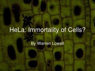
Henrietta Lacks (HeLa) Henrietta Lacks (HeLa) who died in 1951 from cancer of the cervix5
Electrophoretic Mobility Shift Assay (EMSA)6 • apparent molecular weight of DNA-protein complex > unbound DNA • apparent mw of DNA-protein-antibody complex > DNA-protein complex > unbound DNA • used to identify DNA binding proteins
Importance7 • studies have shown that major control of genes and gene expression is done through DNA-protein interactions • e.g.: DNA replication, recombination and repair, transcription, RNA processing, viral assembly • to understand function of interactions, need to know information about structure of DNA-protein complexes, thermodynamics, and kinetics • Eletrophoresis mobility shift assay (EMSA) developed by Garner and Revzin to study these characteristics8.
Conditions of Binding7 • Protein-DNA complexes can be formed by mixing small amounts of protein with labeled DNA in low salt buffer • Formation of complexes can be influenced by many parameters • monovalent ion concentration-low ionic strength (<150mM) increase stability of interaction • presence of non-ionic detergents or carrier proteins-can stabilize product • time and temperature of binding reaction • protein concentration • type and concentration of competitor DNA • Nonspecific competitor such as poly(dA-dT) or poly(dI-dC) used to distinguish between specific and nonspecific binding
Conditions of Native Polyacrylamide Gel7 • mobility of complexes determined by size, charge, and confirmation of protein bound to DNA • composition of gel and electophoretic conditions can alter mobility and stability • higher conc. polyacrylamide stabilizes complex
Applications of EMSA Analysis7 • Quantification • stoichiometric relationships between different complexes • Specificity of protein • DNA binding- perform experiment in presence of increasing amounts of unlabelled competitor • if competitor has high affinity binding site, will compete and decrease visible concentration of detectable complex • Equilibrium constants • obtained by mixing known amount of labeled DNA with increasing conc. of protein and construct standard binding curve • point at which 50% labeled DNA is bound with protein =Keq • Conformational changes of DNA • bent DNA molecules migrate slower than linear (also if bend is in center=slower than on end) • by creating DNA fragments which alter placement of DNA binding site, can study bending activity if protein • Stoichiometric analysis • number of protein that bind per DNA fragment • e.g.- using two different-sized derivatives of same protein-complexes will form three bands (two=homodimers of each derivative + one=heterodimer)
Pros/Cons of EMSA7 • Advantages • don’t need highly purified proteins • can resolve complexes that differ in protein and nucleic acid stoichiometry and/or conformation • easy to separate different species • Disadvantages • no information about sequence of binding site • difficult to adjust all parameters for complete optimization
Procedure • Followed protocol in Peirce EMSA handout2 • Binding Reaction • components: (total volume=20uL) • nuclease free water • 10X Binding Buffer (Tris, KCL, DTT, pH 7.5) • 50% Glycerol • MgCl2 • Poly (dI•dC) (in Tris, EDTA, pH 7.5) • 1% NP-40 • DNA (biotinylated or not) • Protein/lysate • sometimes antibody9 • incubate 20min at room temp • add loading buffer
6% Polyacrylamide gel • 0.5X TBE + 40% acrylamide + APS + TEMED • polymerize 1hr+ • Load/Run gel • use 0.5X TBE buffer • ~200V, 20-25mA, about 20min. • Transfer to (+)nylon membrane • 0.5X TBE, ice cooled • 380mA • 30min. • UV crosslink (5min.)
Block/Wash • Lightshift Blocking Buffer • Lightshift Stabilized Streptavidin-Horseradish Peroxide Conjugate (filtered) • Lightshift 1X Wash Buffer • Lightshift Equilibration Buffer • Detection • Lighshift Luminol/Enhancer Solution • Lightshift Stable Peroxidase Solution • measure chemiluminescene by cooled CCD camera • 5-15min. exposure
Control reaction 1= EBNA control DNA 2=(1)+ EBNA extract 3= (1,2)+ unlabelled EBNA control DNA • Loading dye was omitted from lanes 1 and 2 and therefore they did not have enough glycerol and the DNA diffused away • Still see expected shift in lane 2 due to EBNA DNA-protein complex 1 2 3 experimental results expected results
Control + HeLa cells 1= EBNA control DNA 2=(1)+ EBNA extract 3= (1,2)+ unlabelled EBNA control DNA 4=(1)+ Active Motif HeLa 5=(1)+ Dr. Mascotti HeLa • gel did not run correctly due to buffer dilution error • stopped immediately after lanes entered gel • no shift for EBNA control (lane2) • binding to site for both HeLa samples • binding of approximately same size protein 5 4 3 2 1
Control + HeLa cells (#2) 1= EBNA control DNA 2=(1)+ EBNA extract 3= (1,2)+ unlabelled EBNA control DNA 4=(1)+ Active Motif HeLa 5=(1)+ Dr. Mascotti HeLa • shift in HeLa’s about same size as EBNA shift • could indicate presence of EBNA • or another protein of similar size that recognizes binding site • upper bands= nonspecific binding 1 2 3 4 5
Control + HeLa [DNA] 1= EBNA control DNA 2=(1)+ EBNA extract 3= (1,2)+ unlabelled EBNA control DNA 4=(1)+ Active Motif HeLa 5=(1)+ Dr. Mascotti HeLa • EBNA control shift is missing • could be due to less template available to bind • forgot to put in reaction • Bands in HeLa lanes are the ones that match EBNA shift • most intense bands from previous gel • other bands are gone due to lower concentration of protein 5 4 3 2 1
Preliminary conclusions • EMSA works correctly and detects DNA to at least 1femtomole • Found a few proteins in HeLa cells that recognize EBNA binding site • One of these proteins in each HeLa sample matches shift of EBNA protein-DNA complex • Could be specific or unspecific binding • Could be EBNA or a different protein that happens to have a similar size
Electrophoretic Mobility Shift Assay (EMSA)6 • apparent molecular weight of DNA-protein complex > unbound DNA • apparent mw of DNA-protein-antibody complex > DNA-protein complex > unbound DNA • used to identify DNA binding proteins
Nonspecific Binding and Antibody • none of the bands are as intense as expected based on other labs • other photons make extra spots that don’t have DNA • pockets of substrate between the membrane and saran wrap • contamination that could have gotten some DNA and substrate bound • not sure about the shifts or lanes present to get reliable results
1= biotin-control DNA 2= (1) + extract EBNA 3= (1,2) + unlabeled DNA 4= (1) + Active Motif 5= (1,4) + unlabeled DNA 6= (1,4) + antibody 7= (1) + Dr. M. HeLa 8= (1,7) + unlabeled DNA 9= (1,7) + antibody 10= (1,2) + antibody (control) Possible Interpretation #1 • lane 3- contamination ? (should not have a shift) • bottom of dye front ran off gel • intensities of HeLa inverted from other trials • lane 5- could be a bit of chasing- would indicate specific binding (but not the band that matches EBNA shift) • lane 6- could be a little higher, but is smeared • lanes 9,10- didn’t seem to work at all- low intensity indicates DNA loss
1= biotin-control DNA 2= (1) + extract EBNA 3= (1,2) + unlabeled DNA 4= (1) + Active Motif 5= (1,4) + unlabeled DNA 6= (1,4) + antibody 7= (1) + Dr. M. HeLa 8= (1,7) + unlabeled DNA 9= (1,7) + antibody 10= (1,2) + antibody (control) Possible Interpretation #2 • only see one shift from extracts • unlabeled DNA control still did not work • lane 10- could be supershift from antibody • lane 8,9- still did not work 1 2 3 4 5 6 7 8 9 10
Conclusions • None of the earlier conclusions were disputed • there is binding in the HeLa lysates that match shift with EBNA • could be specific or nonspecific • Antibody could be binding and there is no change in shift due to charge interactions, or conformational changes that counteract the additional weight • Need to run the last experiment again to get reliable results
References • 1. “Crystal Structure of EBNA-1.” Department of Microbiology and Immunology, University of Rochester Medical Center. www.urmc.rochester.edu/smd/mbi/grad2/herp99BB6.html. 2002. (25 April 2002). • 2. “Lightshift Chemiluminescent EMSA Kit.” Pierce. Rockford, IL, 2002. • 3. Solomon, Julie, Carla Fowler, and G. Cooper. “Epstein-Barr Virus.” www.brown.edu/Courses/Bio_160/Projects2000/Herpes/EBV/Epstein-Barr.html. Brown University, 2002. (25 April 2002). • 4. Kang, Myung-Soo, Ciu Chun Hang and Elliot Kieff. “Epstein-Barr virus nuclear antigen 1 activiates transcription from episomal but not integrated DNA and does not alter lymphocyte growth.” Proceedings of the National Academy of Sciences, USA. 98(6), 15233-15238, 2001. • 5. Potier, Beth. Harvard University Gazette. www.news.harvard.edu/gazette/2001/07.19/04-filmmaker.html. President and Fellows of Harvard College, 2002. (25 April 2002). • 6. Lissemore, J. “EMSA.” Molecular Genetics (BL465), John Carroll University, 24 April, 2002. • 7. Norman, Cecilia. “Electrophoresis mobility shift assay (EMSA).” SLU, Uppsala. www.plantae.lu.se/fskolan/arabidopsistexter/CeciliaNorman.html, (30 April 2002). • 8. Garner, M M. Rezvin, A. (1981) Nucl. Acids Res., 9 (13), 3047-3060 • 9. “Mouse Anti-Epstein Barr Virus Nuclear Antigen (EBNA-1) Monoclonal Antibody.” Chemicon International. CA, 2002.
Acknowledgements • Dr. Mascotti • Dr. Lissemore • Pierce • Chemicon International


![Epstein Barr Virus [EBV] - Named after Epstein and Barr](https://cdn5.slideserve.com/9727845/epstein-barr-virus-ebv-named-after-epstein-dt.jpg)