Download
1 / 15
150 likes | 273 Views
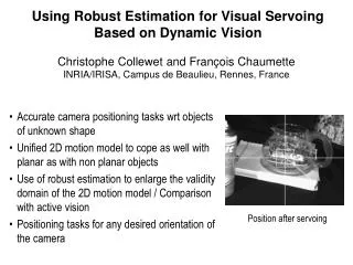
Visual Servoing for Automatic and Uncalibrated Percutaneous Procedures. Ming Li Computer Integrated Surgery II 600.446 2000/3/13. Our Project. Minimally Invasive approach to pelvic osteolysis. cannula. patient. Papers.

E N D
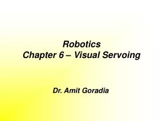
Visual Servoing for Automatic and Uncalibrated Percutaneous Procedures Ming Li Computer Integrated Surgery II 600.446 2000/3/13
Our Project Minimally Invasive approach to pelvic osteolysis cannula patient
Papers Visual Servoing for Automatic and Uncalibrated Percutaneous Procedures M. H. Loser, N. Navab, B. Bascle, R. H. Taylor Proceedings of the Computer Vision and Pattern Recognition (CVPR’00) Motion-Based Robotic Instrument Targeting under C-Arm Fluoroscopy A Patriciu, D Stoianovici, L. L. Whitcomb, T. Jarrett, D. Mazilu, A. Stanimir, I. Iordachita, J. Anderson, R. H. Taylor, L. R. Kavoussi Proceedings of MICCAI’00 A system for Percutaneous Delivery of Treatment with a Fluoroscopically-Guided Robot S. Schreiner, J. H. Anderson, R.H.Taylor, J. Funda, A. Bzostek, A. C. Barnes Proceedings of CVMR and MRCAS1997
Introduction Abstract: Using projective geometry and visual servoing to realize image based approach for semi-automatically guidance of a needle or surgical tool during percutaneous procedures Advantage: 1. No need to pre-operative register 2. No need to use bi-planar X-ray systems 3. No need to calibrate the system prior to use
System Uniplanar X-ray fluoroscope Preoperative procedure: 1. Identify lesion on the X-ray image 2. Choose appropriate insertion point 3. Move robot to place the needle tip at the chosen insertion point
‘ E’ 1 T’ - - 2 1 z Needle pose N1 rotate needle in 1, x y = ’- 0 Method for Uncalibrated Needle Placement Step 1: u Image plane v E T S
Method (cont.) Step 2: Rotate needle around insert point in second plane 2 Needle Pose N2 step1+step2 1
Method (cont.) Step 3: Rotate C-arm to an arbitrary different orientation Move needle in 1 needle pose N3, N3: is the desired 3D pose that aligns the needle with the target in 3D.
Method (cont.) Step 4: Measure the required insertion depth Use 2 marker ball M1, M2
Visual Servo Control Law = ’- < 0.2 Proportional control law: = = f()
Experiment 1.Needle placement with CCD-imaging
Experiment (cont.) 2. Needle placement with X–ray-imaging
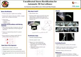
Result Results with CCD-image target: diameter 4mm distance: 60mm Remaining deviation between needle-axis and target after alignment (60 trials) Error in depth estimation after the alignment process Remaining deviation of needle in x’,z’ coordinates after alignment
Result Results with X-ray imaging
Conclusion A simple and accurate method for semi-automatic needle placement, using visual servoing and principles of projective geometry Few Human interaction No stereotactic frame and no prior calibration An alternative to other needle placement technique, which require cumbersome and time consuming calibration procedures