Download

1 / 35
350 likes | 651 Views
In the name of God. Posterior abdominal wall. Dr. Zahiri. consists of : fasciae, muscles and their vessels and spinal nerves. & several viscera : kidneys , suprarenal (adrenal) glands , pancreas , ureters. lumbar vertebrae. five vertebrae between the rib cage and the pelvis.

E N D
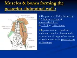
In the name of God Posterior abdominal wall Dr. Zahiri
Dr. Maria Zahiri consists of: fasciae, muscles and their vessels and spinal nerves. & several viscera : kidneys, suprarenal (adrenal) glands, pancreas, ureters
Dr. Maria Zahiri lumbar vertebrae • five vertebrae between the rib cage and the pelvis. • They are the largest segments of the vertebral column The lumbar vertebrae help support the weight of the body, and permit movement
Dr. Maria Zahiri • Iliac fossa • Last ribs
Dr. Maria Zahiri Muscles:PSOAS MAJOR • Morphology: is a long fusiform muscle • Location: on the side of the lumbar region of the vertebral column and brim of the lesser pelvis. • Junction: It joins the iliacus muscle to form the iliopsoas. In less than 50 percent of human subjects,the psoas major is accompanied by the psoas minor.
Dr. Maria Zahiri Muscles:PSOAS MAJOR • Origion: • transverse processes of lumbar vertebrae I-V. • Insertion • lesser trochanter of the femur.
Dr. Maria Zahiri Psoas minor • Origion:the last thoracic and first lumbar vertebrae. • Insertion: iliopectineal eminence.
Dr. Maria Zahiri Iliacus • Origion: iliac fossa on the interior side of the hip bone • Insertion: • lesser trochanter of the femur
Dr. Maria Zahiri Quadratuslumborum Origin: from the lower border of the last rib by four small tendons from the apices of the transverse processes of the upper four lumbar vertebrae Insertion: the internal lip of the iliac crest for about 5 cm.,
Dr. Maria Zahiri diaphragm • is a dome-shaped musculofibrous septum • separates the thoracic from the abdominal cavity, • Its peripheral part consists of muscular fibers that take origin from the circumference of the inferior thoracic aperture and converge to be inserted into a central tendon.
Dr. Maria Zahiri PartOrigin • The muscular fibers may be grouped according to their origins into three parts: • Sternal: two muscular slips from the back of the xiphoid process. • Costal: The inner surfaces of the cartilages and adjacent portions of the lower six ribs on either side, interdigitating with the Transversusabdominis. • Lumbar: Aponeurotic arches, named the lumbocostal arches, and from the lumbar vertebrae by two pillars or crura. There are two lumbocostal arches, a medial and a lateral, on either side.
Dr. Maria Zahiri Crura and central tendon • At their origins the crura(crus) are tendinous in structure • The retrocrural area is the area behind the crus of the diaphragm. • The central tendon of the diaphragm is a thin but strong aponeurosis situated near the center of the vault formed by the muscle
Dr. Maria Zahiri Diaphragm
Dr. Maria Zahiri Blood supply
Dr. Maria Zahiri Thoracolumbar fascia (lumbodorsal fascia • Is a deep investing membrane • covers the deep muscles of the back of the trunk • It is made up of three layers, anterior, middle, and posterior. The anterior layer is the thinnest • The posterior layer is the thickest. • Two spaces are formed between these three layers of the fascia. • Between the anterior and middle layer lies the quadratuslumboru muscle. • The erector spinae muscle is enclosed between the middle and posterior layers.
Dr. Maria Zahiri The posterior layer to the spines of the lumbar and sacral vertebrae The middle layer is attached medially to the tips of the transverse processes of the lumbar vertebrae The anterior layer covers quadratuslumborum
Dr. Maria Zahiri Urinary System Kidneys: Kidneys are retroperitoneal. Lie against posterior abdominal wall on either side of vertebral column. Generally lie adjacent to upper three lumbar vertebrae.
Dr. Maria Zahiri Relation of kidney( ant.)
Dr. Maria Zahiri Relation of kidney( pos.) Diaphragm Transversusabdominis Quadratuslumborum Psoas major
Dr. Maria Zahiri Renal fascia or Gerota's fascia Ant. Layer of renal fascia(fascia of Toldt) lateroconal fascia Pos. Layer of renal fascia(Zuckerkandl's fascia))
Dr. Maria Zahiri kidney Subdivisions: • Cortex. • Medulla with renal pyramids • Pelvis • major and minor calyces (calyx)
Dr. Maria Zahiri Blood supply of kidney • Right and left renal arteries: • Right is longer than the left. • Extrahilar arteries • Right and left renal veins: • Left is longer than the right
Dr. Maria Zahiri Ureter • is a continuation of the pelvis. • Descends retroperitoneally on the anterior surface of the psoas major. • Passes anterior to bifurcation of common iliac.
Dr. Maria Zahiri Blood supply of ureters
Dr. Maria Zahiri Suprarenal (adrenal) glands: Lie against posterior abdominal wall on superior poles of kidneys. Arterial supply: Superior suprarenal arteries: From inferior phrenic artery. Middle suprarenal artery: From abdominal aorta. Inferior suprarenal arteries: From renal artery
Dr. Maria Zahiri Venous drainage: Right suprarenal vein to inferior vena cava. Left suprarenal vein to renal vein.
Dr. Maria Zahiri شاد و سلامت باشید