Download
1 / 66
670 likes | 970 Views
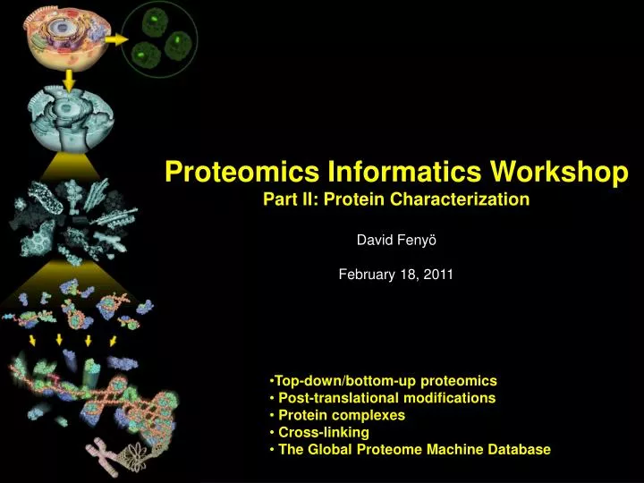
Proteomics Informatics Workshop Part II: Protein Characterization David Fenyö February 18, 2011. Top-down/bottom-up proteomics Post-translational modifications Protein complexes Cross-linking The Global Proteome Machine Database. Proteomics Informatics. MS/MS. Biological System.

E N D
Proteomics Informatics Workshop Part II: Protein Characterization David Fenyö February 18, 2011 • Top-down/bottom-up proteomics • Post-translational modifications • Protein complexes • Cross-linking • The Global Proteome Machine Database
Proteomics Informatics MS/MS Biological System Experimental Design Samples Sample Preparation MS Measurements Data Analysis What does the sample contain? How much? What does the sample contain? How much? Information about each sample Information Integration Information about the biological system
Sample Preparation MS/MS Biological System Experimental Design Enrichment Separation etc Samples Sample Preparation Digestion Top down MS Measurements Bottom up Proteins Peptides Data Analysis What does the sample contain? How much? What does the sample contain? How much? Fragmentation Information about each sample Fragments Information Integration Information about the biological system
Top down / bottom up Top down Bottom up intensity mass/charge
Charge distribution Top down Bottom up 2+ 27+ 31+ 3+ intensity intensity 4+ 1+ mass/charge mass/charge
Isotope distribution Top down Bottom up intensity intensity mass/charge mass/charge
Fragmentation Top down Bottom up Fragmentation
Correlations between modifications Top down Bottom up
Alternative Splicing Top down Exon 1 2 3 Bottom up
Top down Protein mass spectra Fragment mass spectra Kellie et al., Molecular BioSystems 2010
Non-Covalent Protein Complexes Schreiber et al., Nature 2011
Desired Dynamic Range The goal is to identify and characterize all components of a proteome Dynamic Range in Proteomics Distribution of Protein Amounts Number of Proteins Experimental Dynamic Range Log (Protein Amount) Large discrepancy between the experimental dynamic range and the range of amounts of different proteins in a proteome
● Distribution of protein amounts in sample ●# of Proteins in each fraction ●Total amount of peptides that are loaded on column (limited by column loading capacity) ●Total amount of peptides that are loaded on column (limited by column loading capacity) ●Loss of peptides before binding to the column ●# of peptide fractions ●# of peptide fractions ●Loss of peptides after elution off the column ●Distribution of mass spectrometric response for different peptides present at the same amount ●Dynamic range of mass spectrometer ●Detection limit of mass spectrometer Parameters in Simulation
Simulation Results for 1D-LC-MS Tissue No Protein Separation Body Fluid No Protein Separation Complex Mixtures of Proteins Digestion RPC Tissue Protein Separation: 10 fractions Body Fluid Protein Separation: 10 fractions MS Analysis
Success Rate of a Proteomics Experiment Distribution of Protein Amounts Number of Proteins Proteins Detected Log (Protein Amount) DEFINITION: The success rate of a proteomics experiment is defined as the number of proteins detected divided by the total number of proteins in the proteome.
Relative Dynamic Range of a Proteomics Experiment Distribution of Protein Amounts Number of Proteins Proteins Detected RDR90 RDR50 Fraction of Proteins Detected RDR10 Log (Protein Amount) DEFINITION:RELATIVE DYNAMIC RANGE, RDRx, where x is e.g. 10%, 50%, or 90%
Repeat Analysis 2 Analyses 3 Analyses 4 Analyses 5 Analyses 6 Analyses 7 Analyses 8 Analyses 1 Analysis
Repeat Analysis: Comparison of Simulations and Experiments
Tissue Body Fluid 1 1 1 1 2 2 2 2 Tissue Number of Proteins in Mixture RDR50 Success Rate Tissue Body Fluid Body Fluid
Peptide separation Tissue Tissue Tissue Tissue Amount loaded Amount loaded Protein separation Protein separation Protein separation Tissue Tissue Tissue Amount loaded 1. Protein separation 2. Peptide separation 3. Amount loaded Peptide separation Peptide separation Protein separation Protein separation Protein separation Protein separation Amount loaded Peptide separation Amount loaded and peptide separation Order: 1. Protein separation 2. Amount loaded 3. Peptide separation Ranges: Protein separation: 30000 – 3000 proteins in each fraction Amount loaded: 0.1 ug – 10 ug Peptide separation: 100 – 1000 fractions
Localization of modifications Phosphopeptide identification mprecursor = 2000 Da Dmprecursor = 1 Da Dmfragment = 0.5 Da Phosphorylation
Localization of modifications dmin>=3 for 47% of human tryptic peptides Localization (dmin=3) mprecursor = 2000 Da Dmprecursor = 1 Da Dmfragment = 0.5 Da Phosphorylation
Localization of modifications dmin=2 for 33% of human tryptic peptides Localization (dmin=2) mprecursor = 2000 Da Dmprecursor = 1 Da Dmfragment = 0.5 Da Phosphorylation
Localization of modifications dmin=1 for 20% of human tryptic peptides Localization (dmin=1) mprecursor = 2000 Da Dmprecursor = 1 Da Dmfragment = 0.5 Da Phosphorylation
Localization of modifications Localization (d=1*) mprecursor = 2000 Da Dmprecursor = 1 Da Dmfragment = 0.5 Da Phosphorylation
Localization of modifications Peptide with two possible modification sites
Localization of modifications Peptide with two possible modification sites MS/MS spectrum Intensity m/z
Localization of modifications Peptide with two possible modification sites Matching MS/MS spectrum Intensity m/z
Localization of modifications Peptide with two possible modification sites Matching MS/MS spectrum Intensity m/z Which assignmentdoes the data support? 1,1or2, or 1and2?
Visualization of evidence for localization AAYYQK AAYYQK
Visualization of evidence for localization 1 2 3 1 2 3
Estimation of global false localization rate using decoy sites By counting how many times the phosphorylation is localized to amino acids that can not be phosphorylated we can estimate the false localization rate as a function of amino acid frequency. False localization frequency Y Amino acid frequency
How much can we trust a single localization assignment? If we can generate the distribution of scores for assignment 1 when 2 is the correct assignment, it is possible to estimate the probability of obtaining a certain score by chance for a given peptide sequence and MS/MS spectrum assignment.
Is it a mixture or not? If we can generate the distribution of scores for assignment 2 when 1 is the correct assignment, it is possible to estimate the probability of obtaining a certain score by chance for a given peptide sequence and MS/MS spectrum assignment.
Localization of modifications 1and2 1 Ø 1 or 2
Protein Complexes A D A C B Digestion Mass spectrometry
Protein Complexes – specific/non-specific binding Tackett et al. JPR 2005
Protein Complexes – specific/non-specific binding Sowa et al., Cell 2009
Protein Complexes – specific/non-specific binding Choi et al., Nature Methods 2010
Analysis of Non-Covalent Protein Complexes Taverner et al., Acc Chem Res 2008
Determining the architectures ofmacromolecular assemblies Alber et al., Nature 2007
Interaction Partners by Chemical Cross-Linking Protein Complex Chemical Cross-Linking Cross-Linked Protein Complex Enzymatic Digestion MS Proteolytic Peptides Isolation MS/MS Fragmentation Peptides Fragments M/Z
Interaction Sites by Chemical Cross-Linking Protein Complex Chemical Cross-Linking Cross-Linked Protein Complex Enzymatic Digestion MS Proteolytic Peptides Isolation MS/MS Fragmentation Peptides Fragments M/Z
Cross-linking protein n peptides with reactive groups (n-1)n/2 potential ways to cross-link peptides pairwise + many additional uninformative forms Protein A + IgG heavy chain 990 possible peptide pairs Yeast NPC ˜106 possible peptide pairs
Cross-linking Mass spectrometers have a limited dynamic range and it therefore important to limit the number of possible reactions not to dilute the cross-linked peptides. For identification of a cross-linked peptide pair, both peptides have to be sufficiently long and required to give informative fragmentation. High mass accuracy MS/MS is recommended because the spectrum will be a mixture of fragment ions from two peptides. Because the cross-linked peptides are often large, CAD is not ideal, but instead ETD is recommended.