Download
1 / 17
170 likes | 194 Views
The Role of Cilia in Development and Disease. Produced by Gui Ming jie & Li Jing Directed by Pro. Yin. STRUCTURE OF CILIA LEFT–RIGHT PATTERNING ASYMMETRY CILIARY DYSFUNCTION IN DISEASE.

E N D
The Role of Cilia in Development and Disease Produced by Gui Ming jie & Li Jing Directed by Pro. Yin
STRUCTURE OF CILIA LEFT–RIGHT PATTERNING ASYMMETRY CILIARY DYSFUNCTION IN DISEASE
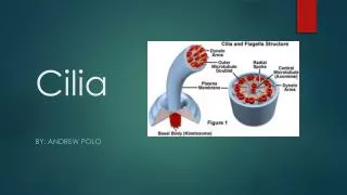
Cilia consist of a basic structureof nine peripheral microtubule doublets arranged around twocentral microtubules (9+2 axoneme). Each outerdoublet is composed of an A and a B tubule (of 13 and 11 protofilamentseach). A central pair of microtubules (C1 and C2), also structurallyand biochemically asymmetric, is present in the center of thering and extends the length of the axoneme. Insome cases the axoneme lacks the central pair apparatus (9+0axoneme). Based on whether the axoneme has a 9+0 or a 9+2 structure,cilia have been defined as primary cilia or motile cilia, respectively.
Recent findings indicate that there are many exceptionsto this definition and favor the distinction into four subtypes: motile 9+2 cilia (e.g. respiratory cilia) motile 9+0 cilia(e.g. nodal cilia) sensory 9+2 cilia (e.g. vestibular cilia) sensory 9+0 cilia (e.g. renal monocilia and photoreceptorconnective cilia)
LEFT–RIGHT PATTERNING ASYMMETRY Cilia are able to generate the currentflow necessary to initiate the signaling cascade for left–rightpatterning in embryos has made an important impact on developmentalbiology.
The ventral surface of the embryonic node inmammals, or of the equivalent structures in other vertebrates, is covered with monocilia that rotate in a clockwise directiongenerating a leftward flow or ‘nodal flow’. When nodal cilia are immotile or absent, nodal flow does notoccur. This leads to randomization of body situs(Fig.2).
The nodal flow and the vortical motion of nodal monocilia indicated with red arrows.
The rotation of the nodal cilia moves the surrounding fluid to the left side of the embryonic midline. It was proposed that the extracellular fluid moved by the nodal cilia contains morphogenetic substances ( i.e., substances that direct embryonic development) that become concentrated on the left side of the embryo, leading to the eventual formation of different organs on different sides of the midline. This proposal is strongly supported by experimental studies in which an artificial flow of fluid across the node could be imposed. When embryos were subjected to a flow of fluid in a direction opposite to that occurring during normal development, the embryo developed with reversed left-right asymmetry.
The first link between cilia and left–right determinationwas suspected by Kartagener who observed that patients withthe heart and abdominal viscera positioned in reversed mirror-image(also called situs inversus) also had respiratory problems,and named that condition Kartagener‘ s syndrome (KS) . Thiscondition is also called primary ciliary dyskinesia (PCD).
CILIARY DYSFUNCTION IN DISEASE Cilia are present in almost all organs of the human body.There is increasing evidence that dysfunction of this largeorganelle is involved in many different human disorders. Sitesof action of cilia that have been implicated in human diseaseare illustrated in Figure 3.
Respiratory cilia PCD, also known as immotile cilia syndrome (ICS)and KS, is characterized by recurrent infectionsof the upper and lower respiratory tract. Motile ciliacovering epithelial cells lining the upper and lower airwaysare responsible for the clearance of the airway . In PCD airway cilia are immotile, dysmotile or absent, whichresults in a reduced mucociliary clearance of the airways. Symptomssuch as respiratory distress, chronic rhinosinusitis andotitis media, persistent cough, and asthma are characteristicof PCD. Often, recurrent infections progress and cause a destructivedilation of the bronchial airway called bronchiectasis .
Cilia of the reproductive system Male infertility due to sperm immotility is frequent in PCD. Female subfertility is less common and is caused by dysfunction of motile cilia from the fallopian tubes and the uterine lining, which are responsible for the oocyte transport. Sperm tails, cilia of the testis efferent ducts and cilia of the female reproductive system share with respiratory cilia the 9+2 ultrastructure
In most PCD patientsultrastructural defects of cilia can be detected by electronmicroscopy. The most common structural defects consistof total or partial absence of dynein arms ( 80%), absence ordislocation of central tubules ( 10%), defects of radial spokes( 6%) and peripheral microtubular abnormalities (3%). PCD represents a heterogeneous group of genetic disorders affecting1/20000 individuals at birth. Inheritance in mostcases is autosomal recessive .
Rare disease manifestations of PCD In a minority of PCD patients the disease is associated withother organ manifestations. hydrocephalus internus eye anomalies such as retinitis pigmentosaand corneal anomalies hearing loss polycystic kidney disorder