Download
1 / 29
290 likes | 723 Views
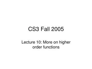
Fall 2005. LAB #11: MAMMALIAN ANATOMY Histology, Fetal Pig Dissection. Histology. The slides you will be examining. Photos by T. Chubb/J. Trout. Epithelial Tissue. Kidney (Simple Cuboidal Epithelium). 40 x. 100x. 400x. Kidney (Simple Cuboidal Epithelium). 40 X. 100 X.

E N D
Fall 2005 LAB #11: MAMMALIAN ANATOMY Histology, Fetal Pig Dissection
Histology The slides you will be examining Photos by T. Chubb/J. Trout
Kidney(Simple Cuboidal Epithelium) 40 x 100x 400x
Kidney(Simple Cuboidal Epithelium) 40 X 100 X This is from a slightly different area of the kidney 400 X
40 X 100 X 400 X EsophagusStratified Squamous Epithelium (non-keratinized)
Centralcanal 100 X osteocyte All the little fine black lines are the caniliculi 400 X Bone
Blood http://www.gihealth.com/images/imgIronDeficiency2.jpg
40 X (ls) Cross section do not use 40 X nucleus 100 X (ls) 400 X (ls) Skeletal Muscle
40 x 100 x nucleus 400 x Cardiac Muscle
Smooth Muscle 100 x 40 x 400 x (ls) 400 x (cs)
Cell body of neuron 100x 400x Nervous Tissue
Dissection: Definition: The process of careful separation for detailed examination & analysis. • TAKE YOUR TIME! • Look BEFORE you cut!
Fetal Pig After external examination, make your initial cuts as shown (follow your lab manual). http://www.ekcsk12.org/science/lelab/incisionsfetalpig.gif
Internal Anatomy Following the lab manual, carefully dissect your specimen. Find all features listed on your “Checklist of Structures”. http://home.satx.rr.com/christenson/Churchill%20Biology/dissec13.jpg
Fetal Heart vs. Adult Heart Is there a difference? Why is there a difference? ….Think about it!
NORMAL HEART (Adult) NORMAL HEART www.mirage-samoyeds.com/ heart.htm
PATENT DUCTUS ARTERIOSIS www.mirage-samoyeds.com/ heart.htm
DEMO: Sheep Heart Dissection External Examination Use the demonstration sheep heart to complete your “Heart Diagram” Handout. http://science.tjc.edu/images/preserved_heart/cut_heart_anterior_label.jpg
DEMO: Sheep Heart Dissection, (cross section view) http://www.sunyniagara.cc.ny.us/val/sheep-heart-3.jpg
DEMO:Sheep Brain(lateral view) http://www.sunyniagara.cc.ny.us/val/sheep-brain-lateral.jpg
DEMO: Sheep Brain (inferior view) http://www.sunyniagara.cc.ny.us/val/sheep-brain-inferior.jpg
TODAY’S PLAN: • Observe histology slides (8). Sketch & label on your “Tissue Types Handout”. (use your lab manual as a guide, Ex. 32). • FetalPigDissection: done in pairs. Follow handout & use your manual, Ex. 33, 34*, 35. Identify all structures on the “Checklist of Structures for the Fetal Pig Dissection”. 3. Observe DEMOs: Sheep Heart, Brain, Shark.
Post-Lab Assignment: To be completed TODAY: (1) histology slide sketches, (2) I.D. pig anatomy, (3) adult heart diagram. To be turned in NEXT WEEK: Lab Manual Questions Exercise 34, pages 573-574.
CLEAN UP! • Dispose of all specimens in the RED BIOHAZARD BAG/BOX provided. • Wash all dissection tools & set out to dry. • Wipe down your lab bench. • Wash your hands before leaving.