Download
1 / 8
80 likes | 91 Views
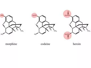
The ventral tegmental area (VTA) is the origin of the mesolimbic dopaminergic system known to play an integral role in mediating reward and development of drug addiction. Although the differences in neuronal plasticity of VTA at various ages remain to be understood, age is known to influence the effects of chronic opioids. In addition, adaptations associated with exposure to opioids within glial populations located in the VTA are poorly understood. The objective of the study was to determine if there are changes in astrocytic immunofluorescent labeling in the VTA following chronic morphine administration in a rat model at different ages: newborn at postnatal day (PD)7 and adult (estimated PD57)

E N D
Research Article http://www.alliedacademies.org/neurology-and-neurorehabilitation-research/ Astrocytic hypertrophy in the rat ventral tegmental area following chronic morphine differs with age. Emily C Goins1, Dusica Bajic1,2* 1Department of Anesthesiology, Critical Care and Pain Medicine, Boston Children’s Hospital, Boston, USA 2Department of Anaesthesia, Harvard Medical School, Boston, USA Abstract The ventral tegmental area (VTA) is the origin of the mesolimbic dopaminergic system known to play an integral role in mediating reward and development of drug addiction. Although the differences in neuronal plasticity of VTA at various ages remain to be understood, age is known to influence the effects of chronic opioids. In addition, adaptations associated with exposure to opioids within glial populations located in the VTA are poorly understood. The objective of the study was to determine if there are changes in astrocytic immunofluorescent labeling in the VTA following chronic morphine administration in a rat model at different ages: newborn at postnatal day (PD)7 and adult (estimated PD57). We hypothesized that increased immunohistochemical labeling of an astrocytic marker, glial fibrillary acidic protein (GFAP) in the VTA following chronic administration of morphine will not differ with age. Two groups of rats were analyzed: chronic morphine and saline control treatment groups. Either morphine (10 mg/kg) or equal volume of saline was given subcutaneously twice daily for 6½ days. On the 7th day of treatment, animals were anesthetized and perfused at one hour after the final drug injection. Coronal sections of the midbrain were processed for immunofluorescent identification of GFAP that was noted at both ages. We report an increase in both (1) GFAP labeling intensity, as well as (2) the percent area occupied by astrocytes that are immunoreactive for GFAP following chronic morphine when compared to saline treatment in the VTA only for the adults (n=6/group) but not infant rats at PD7 (n=5/group). Our findings suggest that adaptations in the mesolimbic dopaminergic system produced by repeated exposure to opioids may be associated with changes in glial function that differ with age. Keywords: Addiction, Astrocytes, Glial Activation, GFAP, Neuroinflammation, Tolerance. Accepted on April 24, 2018 Introduction functions involve providing energy sources and neurotransmitter precursors to neurons, clearing debris, maintaining homeostasis of extracellular ions, and taking up released neurotransmitters to terminate their actions, thereby forming the so-called ‘tripartite synapse’ [13]. Astrocytes also directly participate in synaptic signaling and potentially regulate synaptic plasticity and network excitability [14,15]. Cell membranes of astrocytes bear receptors for many neurotransmitters [16], including opioid peptides [17]. Moreover, astrocytes also exert significant control over the survival of mesolimbic dopaminergic neurons [18] that are part of a well-defined pathway involved in reward processing and addiction [19-22]. This pathway consists primarily of dopamine neurons arising in the ventral tegmental area (VTA) and projecting to the nucleus accumbens. Given that one critical role for astrocytes is the control of NMDA receptor function [23], there is a potential for astrocytes to contribute to the regulation of synaptic plasticity and thereby responses to drugs of abuse in the VTA. Administration of morphine induces reversible hypertrophy and proliferation of astrocytes [24,25]. Such morphological changes, described as activation of glia or astrogliosis, are associated with development of drug dependence [12,26,27]. Finally, pharmacological studies showed that coadministration of a glial inhibitor leads to reduction in the rewarding effects of morphine [28,29]. Although endogenous Opioids, including morphine, relieve acute pain and have become the golden standard for pain treatment in procedural and perioperative settings even for the youngest of patients [1]. However, chronic administration of morphine is associated with negative behavioral effects that, in part, include antinociceptive tolerance and opioid dependence [2]. It was reported that 35%- 57% of infants in neonatal and pediatric intensive care units develop opioid dependence [3]. While the specific mechanisms responsible for analgesic tolerance and dependence remain elusive, it is traditionally thought that chronic morphine administration leads to neuronal plasticity associated with receptor modulation [4-7], and alterations in neuronal excitability [8]. In addition, recent reports implicate enhanced neuroimmune reactivity [9]. Moreover, age has an important effect on the mechanisms of chronic opioids [3]. Despite reports that opioid analgesic abuse has evolved into a national epidemic [10], age differences in cerebral neuroplasticity with age, as well as the ontogeny of specific molecular and cellular mechanisms of chronic morphine administration, remain to be elucidated. Chronic morphine administration directly activates astrocytes [11,12], a form of glia, that are active players in synaptic signaling even under basal conditions. Their ‘housekeeping’ 14 J Neurol Neurorehabil Res 2018 Volume 3 Issue 1
Citation: Goins EC, Bajic D. Astrocytic hypertrophy in the rat ventral tegmental area following chronic morphine differs with age. J Neurol Neurorehabil Res. 2018;3(1):14-21. opioids have been implicated in cell proliferation, migration and differentiation [30], no study looked into the ontogeny of chronic morphine effects on astrocytes. pharmacological groups (split-litter design) [42]. Pups from both sexes were included since gender does not contribute to the degree of antinociceptive tolerance in neonatal rats [43,44]. Furthermore, Craft et al. [45] demonstrated that there are no sex differences in morphine’s antinociceptive effect following 1-week of twice daily 10 mg/kg sc morphine dosing in adult rats. It is poorly understood if chronic morphine administration is associated with adaptations of astrocytes located in the VTA. In this report, we used glial fibrillary acidic protein (GFAP) immunofluorescent analysis to identify qualitative and quantitative changes that might occur following chronic morphine administration in the region of interest, VTA. It is well established that GFAP is an intermediate filament protein specifically expressed in astrocytes [31-35]. To evaluate the age differences of the chronic morphine effect on VTA astrocytes, we analyzed GFAP expression at two different ages of rats: infant at 1-week-old and adult. We hypothesized that astrocytic hypertrophy (astrogliosis) in the VTA following chronic morphine administration in a rat model of antinociceptive tolerance and dependence [36-39] would not differ with age. Brain pharmacological treatment, animals were anesthetized with sodium pentobarbital (100 mg/kg, ip; Hospira Inc., Lake Forest, IL) and perfused through the ascending aorta. Adult animals were perfused with 50 ml of saline, followed by 250 ml of 4% paraformaldehyde in 0.1 M phosphate buffer (PB, pH 7.4, room temperature). Rat pups were perfused with only 100 ml of 4% paraformaldehyde in 0.1 M PB solution. Brains were removed and stored in the same fixative solution overnight (4°C) before cryoprotection in 30% sucrose solution in 0.1 M PB for at least 48 h. Subsequently, brains were frozen and 40 m coronal sections were cut either on a freezing microtome (Leica Microsystems, Wetzlar, Germany; adult brains), or on a cryostat (Leica CM3050S; Leica Microsystems Inc., Buffalo Grove, IL; newborn brains). Although free-floating sections produced better immunolabeling [41], cryostat sections allowed for the better preservation of immature brain tissue being it was more friable in comparison to the adult. Adult brains’ free-floating sections were collected in 0.1 M PB in saline, while pups’ cryostat sections were collected directly on the Superfrost Plus microscope slides (Fisher Scientific, Pittsburg, PA). Finally, pharmacological groups (control and treatment) were matched and brain tissue from different age groups was processed in parallel for immunofluorescent identification of astrocytes. Tissue Fixation and Sectioning. Following Material and Methods Animal Care and Use. Adult male and Pregnant (day 18) Sprague–Dawley rats were derived from Sasco (Charles River Laboratories International, Inc., Wilmington, MA, USA). Adult male animals (250 g; average age postnatal day (PD) 57 according to the supplier) were handled for several days before experiments. Pregnant females were checked at 9 am and 5 pm daily. Pups found at either time were termed 0 days of age and were subsequently used for experiments from PD1-7. All animals were maintained on a 12-h light/dark cycle, and food and water were given ad libitum. The Institutional Animal Care and Use Committee at Boston Children’s Hospital approved the experimental protocols for the use of vertebrate animals in this study. Experiments were conducted according to the U.S. Department of Health and Human Services ‘Public Health Service Policy on Humane Care and Use of Laboratory Animals’ (NIH Publication No. 15-8013, revised 2015) prepared by the National Institutes of Health Office of Laboratory Animal Welfare. All efforts were made to minimize the number of animals used and their discomfort. Immunofluorescence Labeling. To visualize astrocytes, we used immunofluorescent technique against astrocytic marker, glial fibrillary acidic protein (GFAP) as per our previous protocol [41]. Specifically, GFAP immunoreactivity was detected using primary monoclonal mouse anti-GFAP antisera (cat# 3670; Cell Signaling Technology Inc., Beverly, MA) diluted 1:300, and secondary Alexa FluorR 488-conjugated donkey anti-mouse antisera (green fluorescence; cat# A21202, lot# 1022448; Invitrogen Corp., Carlsbad, CA) diluted 1:200. Both primary and secondary antisera were diluted in 0.1 M PB solution with saline, 0.04% bovine serum albumin, 0.3% Triton X-100, and 0.1% sodium azide. Both free floating and brain sections on the slides were incubated in primary antibodies for 2 days at 4°C, and subsequently in secondary antibodies for 1-2 h at room temperature. Cryostat sections were processed on slides in the wet chamber. Sections were rinsed in 0.1 M PB with saline (3 times for 15 minutes) in between and after antibody incubations. Free-floating sections were mounted on slides in 0.05 M PB. Finally, after all the sections were dried, a cover slip was applied with 90% glycerol solution. Immunolabeling with GFAP antisera yielded characteristic staining of astrocytes. Pharmacological Treatment. Morphine sulfate (10 mg/kg; Baxter Healthcare Corp., Deerfield, IL) or equal volume of saline was administered twice daily subcutaneously (sc) for 6½ days. We used a model of repeated morphine exposure that was associated with development of morphine dependence and antinociceptive tolerance in newborn rats [37-39]. These behavioral studies were confirmed in our lab for adult and PD7 rats [36,40]. We used sc injections to minimize nociceptive experience from intraperitoneal drug administration. Detailed description of pharmacological treatment could be found in our recent publications [36,41]. Pharmacological groups included: (1) saline control group, and (2) chronic morphine group (n=6/ group for adult and n=5/group for infant rats). Briefly, animals received twice-daily subcutaneous injections for 6 days (9 AM and 5 PM) and received the last sc injection on the morning of the 7th day. The first day of injections for pups occurred at PD1. Similar to our previous studies of differences in ontogeny of morphine effects [36,41], the progeny from 3 different litters were used. Pups from each litter were assigned to each of the Quantitative analysis The analysis of GFAP immunofluorescence was done using an Olympus IX81 microscope (Olympus America Inc. Melville, NY) equipped with a camera and digital microscopy software (Slidebook v4.2, Olympus). Initially, for each animal, an J Neurol Neurorehabil Res 2018 Volume 3 Issue 1 15
Goins/Bajic average of 3-4 representative coronal sections containing the VTA area [46,47] were photographed bilaterally under 10x magnification at 400 ms exposure (Figure 1). Complementary sets of brain tissue per age were simultaneously processed for immunofluorescent labeling and were subsequently photographed with the same exposure time to minimize inter- assay variability. These images were sufficiently magnified to resolve the GFAP signal. Using the microscopy software (Slidebook v4.2, Olympus), an average labeling intensity of GFAP immunofluorescence per individual astrocyte in the VTA was determined. This was calculated based on analysis of intensity labeling of 5 individual glial cells per picture (total 6-8 pictures/brain). Labeling intensity of the background was also measured and subsequently subtracted from the total intensity of GFAP labeling of each individual glial cell that was analyzed. The average intensity of GFAP immunolabeling/cell ± SD was presented as a percentage (%) from the complementary control processed at the same time, where saline group average values were considered 100%. In addition, an average labeling density was analyzed as total GFAP-labelled surface area/picture ± SD. It was quantified using ImageJ software with a ‘set scale’ of 1.55 pix/micron, and converted the images to grayscale, where 0 represented solid white and 255 ‘threshold’ represented solid black. As a result, the astrocytes in focus on the photograph were solid black so to enhance detection of the edges of the astrocytes (Figure 2). The number of pixels at the threshold point in the frame was calculated by particle analysis and presented as area fraction (% area) of the total pixel area of the image. Average values for both intensity labeling and labeling density (surface area/picture) of GFAP immunoreactivity were compared only to the control values of brains immune processed in parallel. An observer blinded to the treatment group performed the analysis. followed by Tukey post-hoc test. Significance was established at p<0.05. All statistical analyses were done with VassarStats (http://vassarstats.net/), a website for statistical computation. Results In this study, we analyzed both density and intensity of GFAP immunofluorescent labeling in the VTA of adult (estimated PD70), as well as developing rats (at PD7) following chronic morphine treatment. Analyses of GFAP immunofluorescent labeling in VTA were done between 2 groups: saline control and chronic morphine group for each age. Morphological characteristics of GFAP immunofluorescent astrocytes were identified throughout the brainstem at both ages. There were no differences in the individual qualitative appearance of astrocytes between control adult and control developing rats (Figures 2A and 2A’) although they appeared to show distribution difference (viz. more evenly distributed in adult tissue). In control groups, astrocytes had thin and short processes without any clumping or thickening. Qualitative analysis further suggested increased clumping and thickening of astrocytic processes in the area of VTA of adult (n=6/group) but not those of PD7 rats (n=5/group) following prolonged morphine administration (Figure 2B and 2B’). Quantitative analysis confirmed significantly higher % density of GFAP labeling/picture ± SD (F (18,3)=39.08, p<0.0001) both for age (p<0.01), and treatment (p<0.01). With respect to latter, increase in density labeling was noted following chronic morphine treatment (18.08% ± 2.57) in comparison to saline control (11.86% ± 2.99) only in adult rat, as illustrated in Figure 3A. Furthermore, quantitative analysis of the average % intensity of GFAP immunoreactivity/cell ± SD (F(18,3)=5.73, p=0.006) was significant for treatment (p<0.05) but not age. Specifically, significant increase in the intensity labeling following chronic morphine treatment (192.31% ± 85.11) in comparison to saline control (100%) was noted only in adult rats (Figure 3B). In other words, analyses in an infant rat at PD7 (n=5/group) found no differences between saline and chronic morphine treated groups with respect to either % area of GFAP labeling/picture/ brain (6.21% ± 0.51 for saline control; 6.08% ± 1.03 for chronic morphine group) or % intensity of GFAP immunofluorescence/ cell (106.84% ± 16.84 for morphine treatment). Data Analysis Despite differences in tissue sectioning (see above), we controlled for possible differences in the immunofluorescent labeling technique between the two age groups. Thus, for both (1) % intensity of GFAP immunofluorescence/cell and (2) % area of GFAP-labeling/picture in the VTA we used a two-way analysis of variance (ANOVA) with age and treatment as factors, Figure 1. Representative schematics of coronal sections illustrate midbrain at the level of the superior colliculus (SC). Square boundaries indicate the bilateral areas of the ventral tegmental area (VTA) where photomicrographs were taken for analysis. The adult panel (A) corresponds to a plate 44 of the adult rat brain atlas [46], whereas neonatal panel (B) corresponds to a coronal plate 17 (viz. Figure 61) of the neonatal rat brain atlas [47]. The number in the upper right corner of the adult rat coronal section indicates the distance from Bregma. Abbreviations were adapted from the Rat Brain Atlas [46]: Aq, aqueduct; IP, interpeduncular nucleus; ml, medial lemniscus; MG, medial geniculate nucleus; PAG, periaqueductal gray; RN, red nucleus; SC, superior colliculus; SN, substantia nigra. 16 J Neurol Neurorehabil Res 2018 Volume 3 Issue 1
Citation: Goins EC, Bajic D. Astrocytic hypertrophy in the rat ventral tegmental area following chronic morphine differs with age. J Neurol Neurorehabil Res. 2018;3(1):14-21. Discussion In this report, we demonstrate qualitative and quantitative changes related to astrocytic immunolabeling in the rat VTA following chronic morphine administration that differ with age. Qualitative analysis did not appear to show any difference in labeling between control adult and infant rats. Quantitative analysis showed (1) higher density of GFAP-immunofluorescent labeling in the adult tissue at the control conditions, and (2) that chronic morphine administration was associated with increased GFAP immunofluorescence density and intensity in the VTA of adult but not developing rats at 1 week of age. Results suggest that astrocytes could play a role in mediating chronic morphine effects in the VTA that are age-dependent. GFAP labeling with age: Methodological considerations Although GFAP labeling is easily detectable throughout the brainstem as well as the forebrain of the developing rat at 7 days of life [41], presented analysis shows difference in qualitative difference in distribution (Figure 2A and A’), as well as quantitative % surface area of GFAP labeling in the VTA (Figure 3A). As an intermediate filament protein that is expressed in glial cells (astrocytes) [48], its immunoreactivity has been most reliably shown in mature brain with the use of polyclonal antibodies [49]. In contrast, better demonstration of GFAP was shown in immature brain with the use of monoclonal antibodies [49]. One could consider difference in sectioning to contribute to different report in labeling of GFAP in our report being we have also used monoclonal antibodies. Future studies should investigate both PD7 and adult rat labeling following uniform tissue sectioning (e.g. by using cryostat). That would clarify if reported difference in baseline labeling is methodological or inherent to the developmental nature of the immature brain. Other studies so far have only implicated pathophysiology in altered levels of GFAP labeling [50]. Figure 2. Representative photomicrographs of the GFAP immunofluorescent labeling (green) in the VTA of an adult rat (A and B) and infant rat (A' and B') on the postnatal day (PD)7 with different pharmacological treatment: saline control (A and A'), and chronic morphine (B and B'). Black and white panels were created using ImageJ software and illustrate density of GFAP labeling in black. Scale Bar = 50 microm. Chronic morphine and GFAP immunolabeling in VTA with age Adult rat. In the current study, significant increase in labeling intensity as well as density of GFAP immunoreactivity was noted in the VTA of an adult rat treated with chronic morphine. Figure 3. Quantitative analysis of GFAP labeling in the VTA with age and treatment. Graphs summarize (A) average % area of GFAP immunolabeling and (B) average % intensity of GFAP immunolabeling for adult (n=6 sets/12 animals) and PD7 rats (n=5 sets/10 animals) following saline or chronic morphine treatment. Asterisks (*) show significance (p<0.05). A two-way ANOVA with Tukey post-hoc test. J Neurol Neurorehabil Res 2018 Volume 3 Issue 1 17
Goins/Bajic Such increased clumping and thickening of astrocytic processes were described as astrocytic hypertrophy, proliferation and/or astrocytic activation [48]. These results are in accordance with previously published study showing that the long-term systemic morphine administration was associated with a significant increase in GFAP labeling in the VTA [51]. This finding is consistent despite the differences in strain of rats (Wistar instead of Sprague-Dawley), method of chronic morphine administration (pellets instead of twice daily injections for 6½ days), and immunolabeling technique (light microscopy versus immunofluorescence used in our study). Both the Garcia-Perez et al. [51] and our study support original reports of increased GFAP phosphorylation in VTA following chronic morphine administration [52]. Other studies demonstrated hypertrophy of astrocytes in other regions of the mesolimbic reward circuitry: posterior cingulate cortex and hippocampus [12], as well as striatum [53] of adult rats. Although the exact function of morphine-induced astrocytic hypertrophy remains unclear, it is speculated that activated astrocytes may lead to changes in synaptic strength, synaptogenesis and neurogenesis in the brain [54-57]. Furthermore, astrocytic activation (also termed astrogliosis) is considered an important outcome component of the innate immune response that occurs in response to (i) brain injury and neurotoxicity [58,59], (ii) plastic changes associated with increased neuronal activity [59], as well as (iii) following administration of other drugs of abuse [60-62]. of tolerance and dependence to morphine remain controversial [71]. Specifically, opioids activate G-protein-coupled receptors in presynaptic GABAergic neurons. However, neither the pharmacological blockade of these channels, nor the abolition of action potential prevents disinhibition of VTA dopaminergic neurons by morphine [72]. Resent literature implicates presynaptic voltage-activated Ca2+ channels and/ or other components of vesicular trafficking as targets for mu- opioid receptors-mediated inhibition of presynaptic GABA release - as their selective blockade in the VTA disinhibits dopaminergic neurons leading to increased dopamine release in nucleus accumbens [73]. In addition, morphine has been reported to act directly on receptors in astrocytes [19], which constitute the most abundant cell type in mammalian brain [74] that bear receptors for many neurotransmitters [75], including opioid peptides [17], glucose transporters and aquaporin-4 channels for water transport [76,77]. Indeed, recent research supports the view that glial cells, particularly astrocytes, actively contribute to opioid signaling and information processing in the brain [75-81], which has changed the view that astrocytes are nothing more than “sweeping the extracellular milieu free of excess potassium, neurotransmitter, and cellular debris” to facilitate nerve function [82]. Recent study by Shen et al. [83] demonstrated that spinal astrocytic connexin43 contributes to the development of morphine antinociceptive tolerance by activating astrocytes and NMDA receptors, and inhibiting glutamate transporter 1 expression. It is known that drug-induced alterations in synaptic strength in the mesocorticolimbic dopamine system and modulatory glutamatergic neuronal circuits, both part of the brain reward system, underlie drug dependence [84]. Study by Panatier et al. [23] provided evidence that astrocytes are implicated in the regulation of NMDA receptors, typically a receptor for glutamate. Such a contribution of astrocytes to synaptic metaplasticity [24] fuels the emerging concept that astrocytes are dynamic partners of brain signaling. Future studies should elucidate if interaction between the astrocytes and neuronal NMDA receptors is specifically important for morphine dependence in VTA. Infant rat. Although GFAP labeling is easily detectable throughout the brainstem of the developing rat at 7 days of life, chronic morphine administration was not associated with either density or intensity change in GFAP immunofluorescent labeling in the VTA. In the same infant rat model at PD7 age, we previously reported astrocytic hypertrophy associated only with sparse clusters of apoptotic regions of cortex and hippocampus following chronic morphine treatment [41]. Though in vivo studies examining opioid effects on astrocyte physiology during development are limited, in vitro studies have provided some evidence in support of our negative effects. Dose-dependent inhibition of proliferation and induction of morphological differentiation of astrocytes was reported in mixed-glial cell cultures obtained from 1-day-old mice that were treated with chronic morphine [63]. Astrocytes contained the same amount of GFAP immunoreactive intermediate filaments as in control cells after 6 days of morphine treatment. Considering the distinctly unique morphology of glia during early brain development accompanied by significantly higher expression of many cytokines when compared to the adult brain [64], future studies should look into determining the critical period when astrocytes might be mature enough to respond to chronic morphine administration in VTA as described in adult rats. Study by Wu et al. [85] showed that deficiency of aquaporin-4 channels (also known to play a role in chronic opioid effects [86,87] in a knockout mouse attenuates physical dependence that could possibly be mediated by suppression of downregulation of glial glutamate transporter 1 expression. Finally, work by Barr’s group [39,88] (in an infant rat model used in this study) demonstrated that MK-801, NMDA receptor antagonist failed to inhibit morphine withdrawal in the 7-day-old rat but did attenuate the development of morphine dependence in both the 14- and 21-day-old rats. These results suggested that the NMDA receptor is not functionally active in opiate withdrawal until around the second to third week of postnatal life in the rat and that there is a transition period for the NMDA receptor to play a role in the development of opiate dependence and withdrawal. Certainly, future studies should look into the ontogeny of astrocyte-NMDA link with respect to morphine and other drug of abuse effects in the VTA since astrocytes has been shown to have an essential role in glutamatergic neurotransmission and are also affected by morphine [81]. Astrocytes in morphine dependence In the current model, negative behavioral effects of chronic morphine administration such as locomotor activation (proxy of addictive behavior [65-67], and opioid dependence [68], were previously confirmed [37,41]. Opioids are known to activate the main pathway of the brain reward system, mesolymbic dopaminergic system from neurons in the VTA to the nucleus accumbens and prefrontal cortex [69,70]. To date, the molecular and cellular mechanisms mediating the development 18 J Neurol Neurorehabil Res 2018 Volume 3 Issue 1
Citation: Goins EC, Bajic D. Astrocytic hypertrophy in the rat ventral tegmental area following chronic morphine differs with age. J Neurol Neurorehabil Res. 2018;3(1):14-21. Conclusion 9. Johnston IN, Milligan ED, Wieseler-Frank J, et al. A role for proinflammatory cytokines and fractalkine in analgesia, tolerance, and subsequent pain facilitation induced by chronic intrathecal morphine. J Neurosci. 2004;24:7353-65. Presented pilot study supports the hypothesis that chronic morphine administration leads to morphological changes in astrocytes in the VTA, a major reward center of the brain associated with the development of drug dependence that differ with age. A significant increase in density and intensity of GFAP labeling was found in the VTA of adult rats but not in infant rats at 1 week of age following chronic morphine administration. Future studies should look into ontogeny of prolonged opioid effects on astrocytes in VTA, as well as to how described glial changes may alter neuronal function of the reward circuit. Future elucidation of age-specific differences in mechanisms of prolonged opioid dependence will build a foundation for development of age-specific therapies, enhanced safety, and better outcomes for opioid analgesia. 10. Cicero TJ, Inciardi JA, Muñoz A. Trends in abuse of OxyContin® and other opioid analgesics in the United States: 2002-2004. J Pain. 2005;6:662-72. 11. Raghavendra V, Rutkowski MD, DeLeo JA. The role of spinal neuroimmune activation in morphine tolerance/ hyperalgesia in neuropathic and sham-operated rats. J Neurosci. 2002;22:9980-9. 12. Song P, Zhao ZQ. The involvement of glial cells in the development of morphine tolerance. Neurosci Res. 2001;39:281-6. 13. Barres BA. The mystery and magic of glia: A perspective on their roles in health and disease. Neuron. 2008;60:430-40. Acknowledgement Presented work was supported by the Foundation for Anesthesia Education and Research (FAER), Endo Pharmaceuticals, and NIH R03 DA030874 (D. Bajic). Authors would like to thank Allegra L. Spalding, BA for technical assistance with immunofluorescence labeling, as well as Dr. Kathryn G. Commons for providing the equipment and instrumentation to complete the study. The authors do not have any conflict of interest, including specific financial interests, relationships, or affiliations relevant to this article. The content of this article is solely the responsibility of the authors and does not necessarily represent the official views of the NIH. 14. Paixão S, Klein R. Neuron-astrocyte communication and synaptic plasticity. Curr Opin Neurobiol. 2010;20:466-73. 15. Vijayaraghavan S. Glial–neuronal interactions-implications for plasticity and drug addiction. AAPS J. 2009;11:123-32. 16. Haydon PG, Carmignoto G. Astrocyte control of synaptic transmission and neurovascular coupling. Physiol Rev. 2006;86:1009-31. 17. Ridet JL, Privat A, Malhotra SK, et al. Reactive astrocytes: Cellular and molecular cues to biological function. Trends Neurosci. 1997;20:570-7. References 18. Mena MA, de Bernardo S, Casarejos MJ, et al. The role of astroglia on the survival of dopamine neurons. Mol Neurobiol. 2002;25:245-63. 1. Anand KJ. Consensus statement for the prevention and management of pain in the newborn. Arch Pediatr Adolesc Med. 2001;155:173-80. 19. Corrigall WA, Franklin KB, Coen KM, et al. The mesolimbic dopaminergic system is implicated in the reinforcing effects of nicotine. Psychopharmacology. 1992;107:285-9. 2. Jan SA. Introduction: Landscape of opioid dependence. J Manag Care Pharm. 2010;16:S4-8. 3. Anand KJ, Willson DF, Berger J, et al. Tolerance and withdrawal from prolonged opioid use in critically ill children. Pediatrics. 2010;125:1208-25. 20. Phillips PE, Stuber GD, Heien ML, et al. Subsecond dopamine release promotes cocaine seeking. Nature. 2003;422:614-8. 4. Dang VC, Christie MJ. Mechanisms of rapid opioid receptor desensitization, resensitization and tolerance in brain neurons. Br J Pharmacol. 2012;165:1704-16. 21. Salamone JD, Correa M, Mingote SM, et al. Beyond the reward hypothesis: Alternative functions of nucleus accumbens dopamine. Curr Opin Pharmacol. 2005;5:34-41. 5. Martini L, Whistler JL. The role of mu opioid receptor desensitization and endocytosis in morphine tolerance and dependence. Curr Opin Neurobiol. 2007;17:556-64. 22. Wise RA. Dopamine, learning and motivation. Nature reviews. Neuroscience. 2004;5:483-94. 23. Panatier A, Theodosis DT, Mothet JP, et al. Glia-derived D-serine controls NMDA receptor activity and synaptic memory. Cell. 2006;125:775-84. 6. Ueda H, Ueda M. Mechanisms underlying morphine analgesic tolerance and dependence. Front Biosci. 2009;14:5260-72. 24. Abraham WC. Metaplasticity: Tuning synapses and networks for plasticity. Nature Reviews Neuroscience. 2008;9:387. 7. Williams JT, Christie MJ, Manzoni O. Cellular and synaptic adaptations mediating opioid dependence. Physiol Rev. 2001;81:299-343. 25. Narita M, Miyatake M, Shibasaki M, et al. Long-lasting change in brain dynamics induced by methamphetamine: Enhancement of protein kinase C-dependent astrocytic response and behavioral sensitization. J Neurochem. 2005;93:1383-92. 8. Loram LC, Grace PM, Strand KA, et al. Prior exposure to repeated morphine potentiates mechanical allodynia induced by peripheral inflammation and neuropathy. Brain Behav Immun. 2012;26:1256-64. J Neurol Neurorehabil Res 2018 Volume 3 Issue 1 19
Goins/Bajic 26. Miyatake M, Miyagawa K, Mizuo K, et al. Dynamic changes in dopaminergic neurotransmission induced by a low concentration of bisphenol-A in neurones and astrocytes. J Neuroendocrinol. 2006;18:434-44. 43. Thornton SR, Smith FL. Characterization of neonatal rat fentanyl tolerance and dependence. J Pharmacol Exp Ther. 1997;281:514-21. 44. Thornton SR, Wang AF, Smith FL. Characterization of neonatal rat morphine tolerance and dependence. Eur J Pharmacol. 1997;340:161-7. 27. Narita M, Kuzumaki N, Narita M, et al. Chronic pain- induced emotional dysfunction is associated with astrogliosis due to cortical δ-opioid receptor dysfunction. J Neurochem. 2006;97:1369-78. 45. Craft RM, Stratmann JA, Bartok RE, et al. Sex differences in development of morphine tolerance and dependence in the rat. Psychopharmacology. 1999;143:1-7. 28. Narita M, Suzuki M, Kuzumaki N, et al. Implication of activated astrocytes in the development of drug dependence. Ann N Y Acad Sci. 2008;1141:96-104. 46. Paxinos G,Watson C. The rat brain in stereotaxic coordinates. Academic Press. Orlando. FL, USA. 1998. 29. Narita M, Miyatake M, Narita M, et al. Direct evidence of astrocytic modulation in the development of rewarding effects induced by drugs of abuse. Neuropsychopharmacology. 2006;31:2476-88. 47. Ramachandra R, Subramanian T. Atlas of the neonatal rat brain. CRC Press, USA. 2011. 48. Sofroniew MV, Vinters HV. Astrocytes: Biology and pathology. Acta Neuropathol. 2010;119:7-35. 30. Zagon IS, McLaughlin PJ. Increased brain size and cellular content in infant rats treated with an opiate antagonist. Science. 1983;221:1179-80. 49. Sarnat HB, Flores-Sarnat L. Neuropathology of pediatric epilepsy. In: Handbook of clinical neurology. 2013;111:399- 416. 31. Bignami D. Dahl, Astrocyte-specific protein and radial glia in the cerebral cortex of newborn rat. Nature. 1974;252:55-6. 50. Kim R, Healey KL, Sepulveda-Orengo MT, et al. Astroglial correlates of neuropsychiatric disease: From astrocytopathy to astrogliosis. Prog Psychiatry. 2017. 32. Bignami AM, Dahl DO. Specificity of the glial fibrillary acidic protein for astroglia. J Histochem Cytochem. 1977;25:466-9. Neuropsychopharmacol Biol 51. García-Pérez D, Laorden ML, Núñez C, et al. Glial activation and midkine and pleiotrophin transcription in the ventral tegmental area are modulated by morphine administration. J Neuroimmunol. 2014;274:244-8. 33. Bignami A, Eng LF, Dahl D, et al. Localization of the glial fibrillary acidic protein in astrocytes by immunofluorescence. Brain Res. 1972;43:429-35. 34. Eng LF, Ghirnikar RS, Lee YL. Glial fibrillary acidic protein: GFAP-thirty-one years (1969–2000). Neurochem Res. 2000;25:1439-51. 52. BeitnerJohnson D, Guitart X, Nestler EJ. Glial fibrillary acidic protein and the mesolimbic dopamine system: Regulation by chronic morphine and Lewis-Fischer strain differences in the rat ventral tegmental area. J Neurochem. 1993;61:1766-73. 35. Eng L, Vanderhaeghen JJ, Bignami A, et al. An acidic protein isolated from fibrous astrocytes. Brain Res. 1971;28:351-4. 53. Marie-Claire C, Courtin C, Roques BP, et al. Cytoskeletal genes regulation by chronic morphine treatment in rat striatum. Neuropsychopharmacology. 2004;29:2208-15. 36. Bajic D, Berde CB, Commons KG. Periaqueductal gray neuroplasticity following chronic morphine varies with age: Role of oxidative stress. Neuroscience. 2012;226:165-77. 54. Barker AJ, Ullian EM. New roles for astrocytes in developing synaptic circuits. Commun Integr Biol. 2008;1:207-11. 37. Barr GA, Wang S. Tolerance and withdrawal to chronic morphine treatment in the week-old rat pup. Eur J Pharmacol. 1992;215:35-42. 55. Hama H, Hara C, Yamaguchi K, et al. PKC signaling mediates global enhancement of excitatory synaptogenesis in neurons triggered by local contact with astrocytes. Neuron. 2004;41:405-15. 38. Jones KL, Barr GA. Ontogeny of morphine withdrawal in the rat. Behav Neurosci. 1995;109:1189-98. 39. Zhu H, Barr GA. Ontogeny of NMDA receptor-mediated morphine tolerance in the postnatal rat. Pain. 2003;104:437-47. 56. Ullian EM, Christopherson KS, Barres BA. Role for glia in synaptogenesis. Glia. 2004;47:209-16. 40. Bajic D, Soiza-Reilly M, Spalding AL, et al. Endogenous cholinergic neurotransmission contributes to behavioral sensitization to morphine. PloS one. 2015;10:e0117601. 57. Eng LF, Ghirnikar RS. GFAP and astrogliosis. Brain Pathol. 1994;4:229-37. 58. Hill SJ, Barbarese E, McIntosh TK. Regional heterogeneity in the response of astrocytes following traumatic brain injury in the adult rat. J Neuropathol Exp Neurol. 1996;55:1221-9. 41. Bajic D, Commons KG, Soriano SG. Morphine-enhanced apoptosis in selective brain regions of neonatal rats. Int J Dev Neurosci. 2013;31:258-66. 59. Steward O, Torre ER, Tomasulo R, et al. Neuronal activity up-regulates astroglial gene expression. Proc Natl Acad Sci. 1991;88:6819-23. 42. Booze considerations: a determinant of acute neonatal toxicity. Teratology. 1985;31:187-91. RM, Mactutus CF. Experimental design 60. Fattore L, Puddu MC, Picciau S, et al. Astroglial in vivo 20 J Neurol Neurorehabil Res 2018 Volume 3 Issue 1
Citation: Goins EC, Bajic D. Astrocytic hypertrophy in the rat ventral tegmental area following chronic morphine differs with age. J Neurol Neurorehabil Res. 2018;3(1):14-21. response to cocaine in mouse dentate gyrus: a quantitative and qualitative analysis by confocal microscopy. Neuroscience. 2002;110:1-6. 76. Nielsen S, Nagelhus EA, Amiry-Moghaddam M, et al. Specialized membrane domains for water transport in glial cells: High-resolution immunogold cytochemistry of aquaporin-4 in rat brain. J Neurosci. 1997;17:171-80. 61. Hebert MA, O'callaghan JP. Protein phosphorylation cascades associated with methamphetamine-induced glial activation. Ann N Y Acad Sci. 2000;914:238-62. 77. Saadoun S, Papadopoulos MC, Watanabe H, et al. Involvement of aquaporin-4 in astroglial cell migration and glial scar formation. J Cell Sci. 2005;118:5691-8. 62. Itzhak Y, Achat-Mendes C. Methamphetamine and MDMA (ecstasy) neurotoxicity: 'Of mice and men'. IUBMB life. 2004;56:249-55. 78. Volterra A, Meldolesi J. Astrocytes, from brain glue to communication elements: The revolution continues. Nature Reviews. Neuroscience. 2005;6:626-40. 63. Stiene-Martin A, Knapp PE, Martin K, et al. Opioid system diversity in developing neurons, astroglia, and oligodendroglia in the subventricular zone and striatum: Impact on gliogenesis in vivo. Glia. 2001;36:78-88. 79. Beardsley PM, Hauser KF. Glial modulators as potential treatments of psychostimulant abuse. Adv Pharmacol. 2014;69:1-69. 80. Koyama Y. Functional alterations of astrocytes in mental disorders: Pharmacological significance as a drug target. Front Cell Neurosci. 2015;9:261. 64. Schwarz JM, Bilbo SD. The immune system and the developing brain. In: Colloquium Series on the Developing Brain. Morgan & Claypool Life Sciences. 2012: 5-12. 81. Miguel-Hidalgo JJ. The role of glial cells in drug abuse. Curr Drug Abuse Rev. 2009;2:76-82. 65. Joyce EM, Iversen SD. The effect of morphine applied locally to mesencephalic dopamine cell bodies on spontaneous motor activity in the rat. Neurosci Lett. 1979;14:207-12. 82. Diamond JS. Astrocytes put down the broom and pick up the baton. Cell. 2006;125:639-41. 66. Kalivas PW, Stewart J. Dopamine transmission in the initiation and expression of drug-and stress-induced sensitization of motor activity. Brain Res Rev. 1991;16:223-44. 83. Shen N, Mo LQ, Hu F, et al. A novel role of spinal astrocytic connexin 43: Mediating morphine antinociceptive tolerance by activation of NMDA receptors and inhibition of glutamate transporter-1 in rats. CNS Neurosci Ther. 2014;20:728-36. 67. Vezina P, Stewart J. Conditioning and place-specific sensitization of increases in activity induced by morphine in the VTA. Pharmacol Biochem Behav. 1984;20:925-34. 84. Van Huijstee AN, Mansvelder HD. Glutamatergic synaptic plasticity in the mesocorticolimbic system in addiction. Front Cell Neurosci. 2015;8:466. 68. Stein C. Opioids, sensory systems and chronic pain. Eur J Pharmacol. 2013;716:179-87. 85. Wu N, Lu XQ, Yan HT, et al. Aquaporin 4 deficiency modulates morphine pharmacological actions. Neurosci Lett. 2008;448:221-5. 69. Alcaro A, Huber R, Panksepp J. Behavioral functions of the mesolimbic dopaminergic system: An affective neuroethological perspective. Brain Res Rev. 2007;56:283-321. 86. Vitellaro-Zuccarello L, Mazzetti S, et al. Distribution of Aquaporin 4 in rodent spinal cord: Relationship with astrocyte markers and chondroitin sulfate proteoglycans. Glia. 2005;51:148-59. 70. Pierce RC, Kumaresan V. The mesolimbic dopamine system: The final common pathway for the reinforcing effect of drugs of abuse? Neurosci Biobehav Rev. 2006;30:215-38. 87. Zeng XN, Sun XL, Gao L, et al. Aquaporin-4 deficiency down-regulates glutamate uptake and GLT-1 expression in astrocytes. Mol Cell Neurosci. 2007;34:34-9. 71. Al-Hasani R, Bruchas MR. Molecular mechanisms of opioid receptor-dependent signaling and behavior. Anesthesiology. 2011;115:1363-81. 88. Zhu H, Barr GA. Opiate withdrawal during development: Are NMDA receptors indispensable? Trends Pharmacol Sci. 2001;22:404-8. 72. Lowe JD, Bailey CP. Functional selectivity and time- dependence of μ-opioid receptor desensitization at nerve terminals in the mouse ventral tegmental area. Br J Pharmacol. 2015;172:469-81. *Correspondence to: Dusica Bajic Department of Anesthesiology, Critical Care and Pain Medicine, Boston Children's Hospital, Boston, USA, Tel: +1 (617) 355-7737, E-mail: dusica.bajic@childrens.harvard.edu 73. Belardetti F, Ahn S, So K, et al. Block of voltage- gated calcium channels stimulates dopamine efflux in rat mesocorticolimbic system. 2009;56:984-93. Neuropharmacology. 74. Pope A. Neuroglia: Quantitative aspects. Dynamic properties of glial cells, Pergamon Press, London, UK. 1978:13-20. 75. Haydon PG. GLIA: Listening and talking to the synapse. Nature Reviews. Neuroscience. 2001;2:185-93. J Neurol Neurorehabil Res 2018 Volume 3 Issue 1 21