Download
1 / 23
260 likes | 433 Views
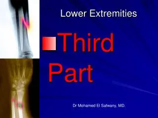
Disturbances of blood and lymph circulation in lower extremities. J. H anáček. Arterial disorders. USG examination of leg arteries. Stenosis of the proximal left Iliac artery. Subclinical stenosis of leg artery. Hemodynamically important stenosis of leg artery.

E N D
Disturbances of blood and lymph circulation in lower extremities J. Hanáček
Stenosis of the proximal left Iliac artery
Analyze mechanisms of 6PS in subjects with chronic occlusive arterial disease Pain Pale Pulseless Paresthesia Paralysis Perishing cold
Case report- The patient is a 18 years old male, who, two months prior to medical consultation, suffered an accidental wound in the middle third of the right thigh.- A brief consultation is done in ambulatory conditions and a tumor is detected in the middle third of the right thigh with a scar in its center.- The formation is incised in ambulatory service and the incision resulted in serious external hemorrhage. Provisional hemostasis is applied by digital pressure in the incision zone; and the patient makes it to the surgical room! He is operated immediately; the axis of the superficial femoral artery is explored and a parietal break is detected (this was the source of the false aneurysm). The parietal breaking is sutured with an enlarging patch and the tissues delimiting the false aneurysm are excised. The evolution is good, uneventful, with no complications, the patient is discharged from the hospital 10 days after the surgery.
A 19-year-old female college student had numbness and the sensation of coldness of her left toes. She had a 3-year smoking history. Gangrene of the left foot developed rapidly. Angiography revealed peripheral arterial occlusion of both legs and arms. Detailed laboratory examination excluded collagen disease, a hypercoagulable state, and juvenile atherosclerosis. Below-knee amputation of the left leg was performed. Typical histologic findings of Buerger’s disease were observed in the crural arteries and saphenous veins. The clinical course was uneventful after the patient stopped smoking. This is the second case report of Buerger’s disease in a woman in the second decade of life. It is important that a correct diagnosis of Buerger’s disease be established, because the disease process is benign, compared with collagen disease, if the patient stops smoking.
48 years male, with DM type 1 (30 years duration), hypertension, hyperuricaemia, and BMI 32 suffers from intermittent claudicating, leg pain, and a wound (skin defect) on the right leg resistant to treatment Analyze how factors describedbelowmay contribute to occlusive arterial disease - Diabetes mellitus - Hyperglycaemia - Hypertension - Hyperuricaemia - BMI 32 How this situation can be promoted by microangiopathy and neuropathy? Is age 48 typical for having advanced occlusive arterial disease? Explain.
Deep venous trombosis Risk factors - acquired
Inherited • Antithrombin deficiency[8] • Protein C deficiency[8] • Protein S deficiency (type I)[11] • Factor V Leiden[e] • Prothrombin G20210A • Dysfibrinogenemia[19] • Non-O blood type • Mixed • Low free protein S[11] • Activated protein C resistance[11] • High factor VIII levels[31] • Hyperhomocysteinemia[8] • High fibrinogen levels[8] • High factor IX levels[8] • High factor XI levels[8]
Case report 70 yr old female is complaining about leg pain, mainly after exercise, not during it, she describes her feeling as heavy legs, especially at the evening. Physical examination showed edematous ankles, brown color stains on the skin around the ankles, skin lesion, resistant to treatment. She does not have DM, hypertension, but she had varices for more than 40 years. VF on admission HR 70 bpm/, BP 160/100 mmHg, BR 16 bpm. - Define chronic venous insufficiency - Explain mechanisms leading to clinical presentation of this patient - What additional signs and symptoms would you expect? - Explain relationships between varices, DVT and development of chronic venous insufficiency - Why she suffers from skin trophic changes instead she has rather venous than arterial disease? - Explain mechanism of diffusion hypoxia - Explain mechanisms od edema formation in this case