Download
1 / 26
370 likes | 933 Views
Infectious Esophagitis. Immunocompromised Host -Steroids, Chemo/Rad therapy, AIDS, Transplant patients Endoscopic Appearance Location - Often more proximal than reflux. Candidal Esophagitis. Normal Flora, ubiquitous agent

E N D
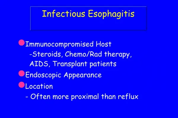
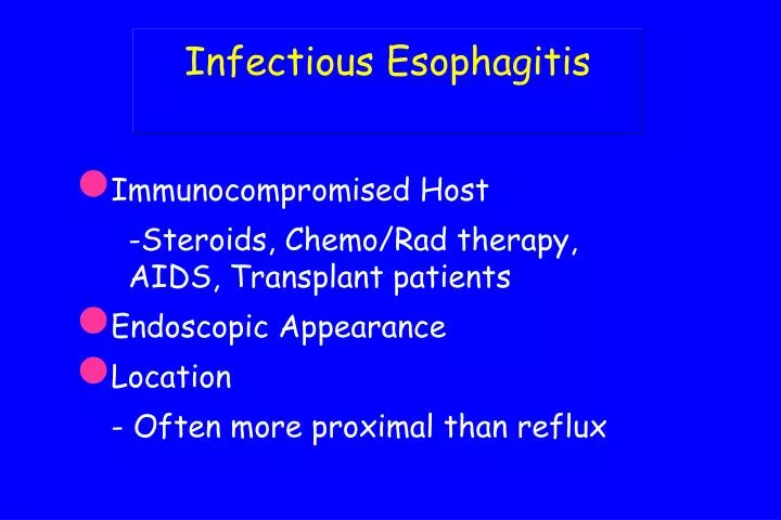
Infectious Esophagitis • Immunocompromised Host • -Steroids, Chemo/Rad therapy, AIDS, Transplant patients • Endoscopic Appearance • Location • - Often more proximal than reflux
Candidal Esophagitis • Normal Flora, ubiquitous agent • - may gain selective advantage after antibiotics or in immunocompromised • Acute presentation of odynophagia/dysphagia • Endoscopic appearance of white -yellow plaques - “cottage cheese”
Candidal EsophagitisHistopathology • Clumps of necrotic squamous debris • Neutrophils in surface epithelium • - Sometimes large aggregates of lymphocytes • Pseudohyphae grow perpendicular to axis of superficial squamous cells • PAS or GMS stains help identify organism
Candida PAS stain
Candida Lymphocytic reaction
Herpes Esophagitis • Either Herpes Simplex type 1 or 2 • Reactivation in immunocompromised adults • - usually type 1 • Neonates - esophagus involved by disseminated intrapartum infection • - usually type 2
Herpes Esophagitis • Acute presentation of odynophagia/dysphagia, may have GI bleeding • Endoscopic appearance of grouped vesicles, erosions, or ulcers - depending on stage • Located in mid to lower 1/3 of esophagus
Herpes EsophagitisHistopathology • Viral inclusions in squamous epithelium • - Cowdry A and B inclusions • Multinucleated cells with smudgy nuclear inclusions • Aggregates of macrophages in exudate
HSV Macrophages often seen under infected epithelium
CMV Esophagitis • Reactivation in immunocompromised hosts • - AIDS and Transplant patients at high risk • Accompanied by systemic infection • - unlike HSV • Clinical presentation identical to HSV • Single distal ulcer most common endoscopic appearance
CMV EsophagitisHistopathology • Nuclear and cytoplasmic inclusions present in endothelial cells, macrophages, smooth muscle / stromal cells - not present in squamous cells • Nuclear inclusion is classically Cowdry type A • Cytoplasm of cell may show granular inclusions, but these form after nuclear inclusions and may not be present in small biopsy specimens
HIV Associated Esophagitis • Giant esophageal ulcers for which no pathogen can be found • - Deep ulcers in mid or distal esophagus, often greater than 1 cm in diameter • - HIV RNA present by in-situ studies • - Treatment with steroids is helpful, but patients often relapse after steroids are withdrawn
Where to Biopsy? • In Candida, the superficial necrotic debris is most likely to have the diagnostic yeast and pseudohyphae • In HSV, the edge of the ulcer is most likely to harbor inclusions • In CMV, the granulation tissue and muscle from the deepest portion of the ulcer are probably best