Download

1 / 91
930 likes | 1.3k Views
Neuromuscular Fatigue. Muscle Physiology 420:289. Agenda. Introduction Central fatigue Peripheral fatigue Biochemistry of fatigue Recovery. Introduction Fatigue. Common definition: Any reduction in physical or mental performance Physiological definitions:

E N D
Neuromuscular Fatigue Muscle Physiology 420:289
Agenda • Introduction • Central fatigue • Peripheral fatigue • Biochemistry of fatigue • Recovery
Introduction Fatigue • Common definition: • Any reduction in physical or mental performance • Physiological definitions: • The gradual increase in effort needed to maintain a constant outcome • The failure to maintain the required or expected outcome/task
Mechanisms Difficult to Study • Many potential sites of fatigue • Task specificity • Central vs. peripheral factors • Environment • Depletion vs. accumulation • Interactive nature of mechanisms • Compartmentalization • Training status
Potential Outcomes of Fatigue • Muscle force: Isometric or dynamic • Peak force and RFD reduced • Rate of relaxation: • Reduced Figure 15.6, McIntosh et al., 2005 Adopted from Garland et al. (1988)
Potential Outcomes of Fatigue • Muscle velocity and power: • Peak and mean reduced McIntosh et al., 2005
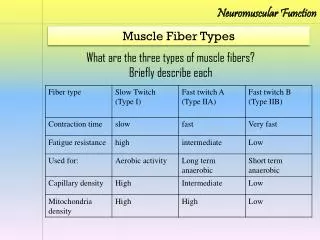
Potential Outcomes of Fatigue • EMG • Increases with fatigue (submaximal load) as CNS attempts to recruit more motor units • Power frequency spectrum shifts to left • FT MUs fatigue resulting in greater stimulation of ST MUs (lower threshold lower frequency)
Potential Outcomes of Fatigue • Ratings of perceived exertion • Rate of fatigue • Fatigue index Wingate • Time to fatigue
Mechanisms of Fatigue • Fatigue can be classified in many ways: • Psychological vs. physiological • Neuromuscular vs. metabolic • Central vs. peripheral
Agenda • Introduction • Central fatigue • Peripheral fatigue • Biochemistry of fatigue • Recovery
Central Fatigue - Introduction • Central fatigue: A progressive reduction in voluntary activation of muscle during exercise • Difficult to study however strong indirect evidence
Central Fatigue - Introduction • Central fatigue may manifest itself in several ways: • Emotions and other psychological factors • Afferent input (pain, metabolites, ischemia, muscle pressure/stretching) • Intrinsic changes of the neuron (hyperpolarization of RMP)
Bottom line: Central fatigue causes neural inhibition greater voluntary effort to drive any motor unit Figure 1, Kalmer & Cafarelli, 2004
Evidence of Central Fatigue • Reduced motor unit firing rate and ½ relaxation time • Suggests less central drive
Evidence of Central Fatigue • Concept of muscle wisdom • Decline in MU firing rate does not correlate well with decline in force • As MU firing rate declines ½ relaxation time increases (prolonged contractile mechanism) • Prolongation steady force maintained with lower MU firing rate • Increased efficiency? • Eventual fatigue is imminent
Evidence of Central Fatigue • Best evidence: Improvement in performance with severe fatigue • Sudden encouragement • Last “kick” at end of race
Agenda • Introduction • Central fatigue • Peripheral fatigue • Biochemistry of fatigue • Recovery
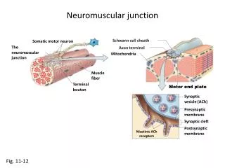
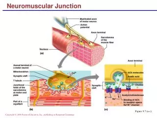
Peripheral Fatigue • Potential sites include (but not limited to): • Impulse conduction of efferent neurons and terminals • Impulse conduction of muscle fibers • Excitation contraction coupling • Sliding of filaments
Efferent Neurons and Terminals • Impulse conduction may fail at branch points of motor axons • Unusual Branch point diameter < axon diameter • Evidence: Krnjevic & Miledi (1958) Zhou & Shui, 2001
Krnjevic & Miledi (1958) • Rat diaphragm motor nerve • Motor end plates of two fibers within same motor unit observed • Fatigue One fiber did not demonstrate motor end plate depolarization with stimulation • Conclusion: Branch point failure
Efferent Neurons and Terminals • Note: “Dropping out” of muscle fibers in single muscle fiber EMG studies is very rare • More research is needed
Efferent Neurons and Terminals • ACh release from axon terminals? • ACh is synthesized and repackaged quickly even during repetitive activity • Safety margin: Very little ACh is required to stimulate AP along sarcolemma • At least 100 vescicles released/impulse • Not considered a site of peripheral fatigue
Peripheral Fatigue • Potential sites include (but not limited to): • Impulse conduction of efferent neurons and terminals • Impulse conduction of muscle fibers • Excitation contraction coupling • Sliding of filaments
Impulse Conduction Muscle Fibers • The ability of the sarcolemma to propagate APs will eventually fail during repetitive voluntary muscle actions • Attenuation is modest • Mechanism: Leaking of K+ from cell hyperpolarization of RMP
Figure 15.6, McIntosh et al., 2005 Adopted from Garland et al. (1988)
Peripheral Fatigue • Potential sites include (but not limited to): • Impulse conduction of efferent neurons and terminals • Impulse conduction of muscle fibers • Excitation contraction coupling • Sliding of filaments
Excitation-Contraction Coupling • Potential sites of fatigue: • Tubular system: • T-tubules • Sarcoplasmic reticulum
ECC Fatigue T-Tubules • Mechanism: • Inability of AP to be propagated down t-tubule • Due to pooling of K+ in t-tubule (interstitial fluid) • Recall: • Muscle activation causes: • Increase of intracellular [Na+] • Decrease of intracellular [K+] • Na+/K+ pump attempts to restore resting [Na+/K+] • Na+/K+ pump is facilitated by: • Increased intracellular [Na+} • Catecholamines
ECC Fatigue T-Tubules • T-tubule membrane surface area is small • Less absolute Na+/K+ pumps • Pooling of K+ in t-tubules hyperpolarizes t-tubule RMP • Time constant for movement of K+ from t-tubules = ~ 1s • Does 1 s of rest alleviate fatigue? • More mechanisms!
ECC Fatigue Sarcoplasmic Reticulum • Several potential mechanisms: • Impaired SERCA function • Reduced uptake of Ca2+ prolonged relaxation? • Impaired RYR channel function • Reduced release of Ca2+ less crossbridges? • General rise in intracellular Ca2+ • Increased uptake of Ca2+ by mitochondria reduced mitochondrial efficiency?
Peripheral Fatigue • Potential sites include (but not limited to): • Impulse conduction of efferent neurons and terminals • Impulse conduction of muscle fibers • Excitation contraction coupling • Sliding of filaments
Sliding of Filaments • Troponin: Two potential mechanisms of fatigue • Decreased responsiveness: Less force at any given [Ca2+] • Decreased sensitivity: More [Ca2+] needed for any given force
Agenda • Introduction • Central fatigue • Peripheral fatigue • Biochemistry of fatigue • Recovery
Biochemistry of Fatigue • Metabolic fatigue • Depletion • Accumulation • Metabolic depletion and accumulation is related to central and peripheral fatigue
Metabolic Depletion • Phosphagens • Glycogen • Blood glucose
Phosphagen Depletion • Phosphagens include: • ATP • Creatine phosphate CP is most immediate source of ATP due to creatine kinase Rate of ATP: High Capacity: Low
Phosphagen Depletion • Pattern of CP/ATP depletion • CP and ATP deplete rapidly • CP continues to deplete task failure • ATP levels off and is preserved
Phosphagen Depletion • ATP depleted why task failure? • Down regulation of “non essential” ATP utilizing functions in order to maintain “essential” functions • Free energy theory
Phosphagen Depletion • Bottom line: • CP depletion results in fatigue during high intensity exercise • CP supplementation delays onset of task failure