Download
1 / 17
170 likes | 173 Views
PHYLUM PORIFERA. Level of body organization? Symmetry? Name of Middle layer? = Acellular matrix - location of structural elements & has cells moving through it. Name the structural elements. Which components are used to ID sponges? Name the moving cells. Form of locomotion?

E N D
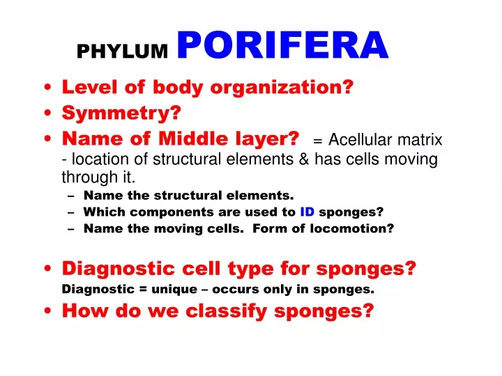
PHYLUM PORIFERA • Level of body organization? • Symmetry? • Name of Middle layer?=Acellular matrix - location of structural elements & has cells moving through it. • Name the structural elements. • Which components are used to ID sponges? • Name the moving cells. Form of locomotion? • Diagnostic cell type for sponges? Diagnostic = unique – occurs only in sponges. • How do we classify sponges?
PHYLUMPORIFERA • CELLULAR level of body organization • ASYMMETRICAL(entire body) or RADIAL (not perfect) • Middle layer = MESOHYL Spongin (a collagen protein) & Spicules Spicules (Ca or Si) are used to ID sponges Calcarea (Ca) Demospongiae (Ca &/or Si) Hexactinellida (Si) Amoebocytes = archaeocytes are amoeboid • Diagnostic cell type: CHOANOCYTE = flagellated collar cell. (Collar cells exist in other phlya but they are not flagellated.)
Classification of sponges is byBODY TYPE Syconoid = middle-sized Asconoid = smallest TYPES are not taxa but basic groups …based on their internal architecture …i.e. the location of their WHAT? Leuconoid = Largest
PHYLUM Porifera TYPE ? In the jar, these sponge specimens look like white or transparent plant roots.. In lab you could only look at a whole specimen (as above) in a jar or at prepared slides.
PHYLUM Porifera TYPE Asconoid NOTE: Many of our slide specimens have been stained red or green. (Look like……..??????) This is the smallest and simplest sponge type. (i.e. they are too small to dissect.) Name often used for this most unit?
PHYLUM Porifera TYPE Asconoid BSU – Basic Sponge Unit. Choanocytes are located in the spongocoel. What is the function of a gemmule?
PHYLUM Porifera TYPE ? What is this? Name this aperture? What is this?
PHYLUM Porifera TYPE Asconoid Gonad Long spicules at osculum neck Bud Terms you need to know:spicules, spongocoel, bud & osculum.Compare to fig 1.3-A in your lab manuals.
Incurrent Pores (Ostia), Porocytes and Prosopyles • Incurrent pores or ostia are the openings through which water first enters a sponge. These are usually formed by several cells. • The PROSOPYLE is the name given to the entryway (pore) leading into the area of choanocytes. It is formed by one donut- shaped cell, the porocyte.
Asconoid Sponges As an incurrent pore or ostium, this opening brings water directly into the sponge. (BLACK) It also serves as a prosopyle, (BLUE) bringing water into contact with the choanocytes lining the spongocoel. Thus it has a dual function, serving as the incurrent pore or ostium and as a prosopyle. The actual opening is formed by a single cell, the porocyte.
Syconoid Sponges The ostia/incurrent pores in syconoid sponges are generally made of several cells (pinacocytes). (DOTTED BLACK) Water enters the sponge through this entryway and moves into the incurrent canal. Water leaves the incurrent canal area to enter the radial canal (area of choanocytes) via the prosopyle – (a porocyte cell) Water leaves the area of choanocytesvia a much larger pore, made by many cells = the apopyle.
PHYLUM Porifera TYPE Syconoid Note the prominent spicules Sycons (Syconoid sponges) are the ‘middle-sized’ sponges. Their choanocytes are located in the ? canals.
O Ostia I A R (rough walls) S I (slick walls) P S PHYLUM Porifera TYPE Syconoid l.s. & c.s. views l.s. I I R R Water flow:Ostium -> Incurrent canal (I) -> Prosopyle channel (P) (a porocyte) ->Radial canals (R) (area of choanocytes) -> Apopyle channel (A) -> Spongocoel (S) -> Osculum (O)
Phylum? Class? Choanocytes are located where?
PHYLUM PoriferaTYPE Leuconoid No classes! TYPES! Leucons/Leuconoid sponges the most complex. Choanocytes are located in flagellated chambers.Any large sponge is most likely a leuconoid - type sponge.
Leuconoid Sponges The ostia (several cells) allow water to enter incurrent canals. Water leaves these to enter the flagellated chambers (area of choanocytes) via the prosopyles (porocytes)
ASEXUAL Marine Budding Fragmentation Regeneration Freshwater sponges Gemmules + 3 methods above SEXUAL Male & female gametes are formed. Archeocytes eggs Choanocytes sperm Fertilization is involved. Planktonic larvae or mini flagellated colonies are released to colonize newareas. Sponge Reproduction Sponges are usually monoecious but can be dioecious