Download
1 / 37
380 likes | 625 Views
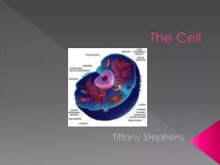
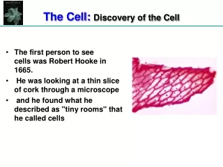
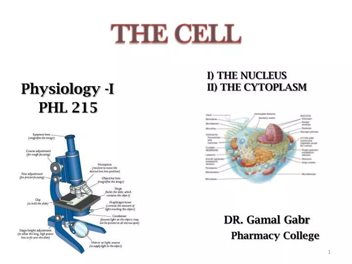
THE CELL. Physiology -I PHL 215. DR . Gamal Gabr Pharmacy College. I) THE NUCLEUS II) THE CYTOPLASM. Cell theory. Cells are measured using the unit of micrometer (µm). 1 millimetre (mm) = 1000 µm. 1 µm = 1/1000 mm. 1- All living organisms are made of one or more cells .

E N D
THE CELL Physiology -I PHL 215 DR. GamalGabr Pharmacy College I) THE NUCLEUSII) THE CYTOPLASM
Cell theory • Cells are measured using the unit of micrometer (µm). 1 millimetre (mm) = 1000 µm. 1 µm = 1/1000 mm. 1- All living organisms are made of one or more cells. 2- Cell is the smallest unit of the living organisms that is capable of performing life functions. 3- All cells come from pre-existing cells through cell division. • NOTES: 1- Living organisms may be: • Unicellular organisms: Organisms that are made of only one cell. • Multicellular organisms: Organisms that are made of many cells.
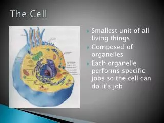
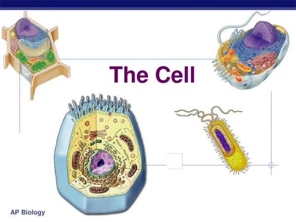
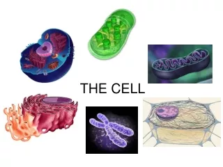
The Cell • Definition: Cell is the smallest unit of the living organisms that is capable of performing life functions. • Types of cells: 2 types 1- Prokaryotic cells (Prokaryotes). 2- Eukaryotic cells (Eukaryotes). • Both types of cells have many things in common as: 1- Cell membrane (Plasma membrane) 2- Cytoplasm 3- Genetic material
Prokaryotic cells • Cells that do not has a nucleus or any other membrane-bound organelles. • Prokaryotic cells are much smaller than eukaryotic cells • Example: Unicellular organisms as Bacteria Eukaryotic cells • Cells that contain nucleus and other membrane-bound organelles. • Unlike prokaryotes, eukaryotic organisms may be unicellular as in amoebae or multicellular as in plants and animals. Animal cell Plant cell
The Eukaryotic Cell – Animal Cell • The human body is made up of smaller structures. • These structures are of 4 major types: 1- Cells • Cell is the smallest unit of the living organisms that is capable of performing life functions. 2- Tissues • A tissue is composed of many similar cells. 3- Organs • An organ is composed of different types of tissues to perform a specific function. 4- Systems • A system is composed of different types of organs that arranged together to perform complex functions for the body.
THE CELL • Animal cells are oval to square structures usually containing nucleus, cytoplasm and enclosing plasma membrane. • Size • Most cells = 1 - 100 µm So cells are seen only under the microscope. • Number: • The entire body contain 100 trillion cells of different types. Trillion = 1,000,000,000,000. • Each type of cells is adapted to perform one particular function: • Nerve cells Sensation • Muscle cells Motility • Gland cells Secretion
The internal environment of the body • The internal and external environment of a cell is mostly water. • This environment (water) contains charged particles like sodium and potassium ions, simple sugars like glucose, amino acids, and other molecules that are useful to the cell. • Typical volume of water in the body = about 42 liters • This volume represents 60% of adult body weight adult body weight = 70 kg • Water carries nutrients to all parts of the body so called body fluid.
Body fluids: 1- Intracellular fluid (ICF) • Found inside the cells. • Typical volume : about 28 liters (2/3 of body fluid). NOTE: • Any material that is found inside the cell is considered to be intracellular 2- Extracellular fluid (ECF). • Found outside the cells. • Typical volume:about 14 liters (1/3 of body fluid). • Composed of: a- Interstitial fluid (between cells). b- Plasma (in blood). NOTE: • Any material that is found outside the cell membrane of a cell is considered to be extracellular
a- Interstitial fluid (tissue fluid) • Itis a fluid between the cells. • Typical volume:about 11 Liters, so it is the main component of the ECF. • Contain amino acids, sugars, fatty acids, enzymes, hormones, salts. b- Plasma Typical volume:3 Liters ECFmoves throughout the body in a constant motion • Rapidly transported in the circulating blood • Diffuse through capillary walls • Connects between blood and ICF ► So, all cells live in the same environment i.e the ECF represents the internal environment of the body.
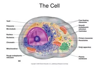
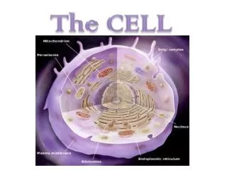
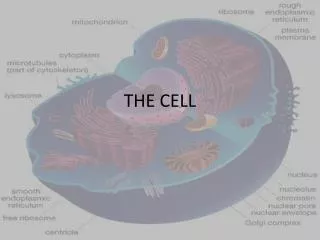
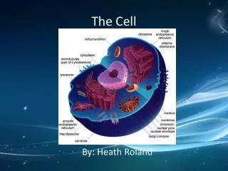
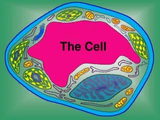
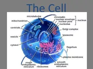
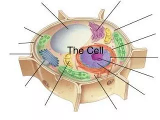
Cell Structure & Function Typical cell composed of3 major parts: ❶ Nucleus ❷ Cytoplasm ❸ Plasma membrane. (Cell membrane)
❶ The Nucleus • The nucleus is a spherical-shaped organelle present in every eukaryotic cell. • Nucleus is the largest structure in the cell as it represent 10 % of the total cell volume. • It contains the genetic material (DNA) • The nucleus is the Control center of the cell as it regulates all cell activities by controlling the cellular enzymes. The Nucleus consists of: I. Nuclear envelope II. Nucleoplasm: a) Chromatin b) Nucleolus
I. Nuclear Envelope: • Separates the nucleus from other parts of the cell. • It composed of 2 layers (inner & outer layers): • The outer layer: • Connected to the endoplasmic reticulum (ER). • Contain pores (nuclear pores) to facilitate the exchange between nucleus & cytoplasm. Function: 1- Protect the nucleus 2- Control movement of particles in and out the nucleus.
II. The Nucleoplasm: • Highly viscous liquid • Composed of chromatin & nucleolus. A- Chromatin: • The chromatin is composed of DNA (deoxyribonucleic acid). • At the time of cell division chromatin arrange in groups called Chromosomes. NOTES: • Chromosomes are the genetic material of the cell. • RBC's do not have chromosomes because they do not have nucleus
Number of chromosomes varies in different species: • Horse 38 • Human 46 • Chromosomes occur in pairs 23 pairs in human. Pass to a new individual B- Nucleolus: • Spherical-shaped large amount of proteins & rRNA (ribosomal RNA). • Function of the nucleolus: Synthesis of ribosomes which are responsible for protein synthesis
Functions of the Nucleus: 1- Control all cell activities (by controlling the cellular enzymes.). 2- Store the hereditary material DNA 3- Produce ribosomes Protein synthesis.
❷ The Cytoplasm • It is clear jelly-like material that fill the area between plasma membrane and nuclear envelope. • It composed of Cytosol and Organelles A. Cytosol: • Cytosol (the intracellular fluid) is the semifluid portion of cytoplasm. • It composed mainly of water that contains inclusions (proteins, lipids, carbohydrates), dissolved salts, gases, and enzymes. Functions of Cytosol: • Suspension of all organelles in the cell. • Maintains the shape and consistency of the cell. • It is a storage place for substances essential to life.
B. Organelles: Organelles means Little organs. • They are small structures found within the cytosol. • Each organelle has a characteristic shape and perform specific function for the cell. There are 2 types of organelles: I) Membrane Bound Organelles 1- Endoplasmic Reticulum (ER) 2- Golgi apparatus 3- Mitochondria 4- Lysosomes, Peroxisomes, Secretory Vesicles II) Non-membrane Bound Organelles 1- Ribosomes 2- Cytoskeleton
I) Membrane Bound Organelles 1- Endoplasmic Reticulum (ER) • The ER is a network of flattened sacs and tubules. • The space inside of the ER is called the lumen. • The ER extends from the plasma membrane to the nuclear envelope. • The main function of the ER is to transport materials through the cell, So, the ER represent the transport system of the cell. • There are 2 types of the ER that differ in both structure and function: A- Rough ER B- Smooth ER • Cells specializing in the production of proteins have a larger amount of Rough ER while cells producing lipids have a greater amount of Smooth ER.
A- The Rough ER • It is called Rough ER because its surface is covered with Ribosomes. • Ribosomes give the ER its "rough“ appearance. • Ribosomes are responsible for protein synthesis. • Typically, the Rough ER is a network of flattened sacs. • The rough and smooth ER are usually interconnected and the proteins made by the rough ER move into the smooth ER to be transferred to other locations.
In the Rough ER, ribosomes synthesize the protein (not mature). • Then, a part of the Rough ER membrane breaks off to form a vesicle around the protein. The vesicle is considered as the transport vehicle for the protein. • The vesicles transport protein to Golgi apparatus for further processing and modification (in case of proteins which are not mature) OR moves directly to the plasma membrane (in case of proteins which are mature) . Functions of the Rough ER: Synthesis of proteins Examples of protein synthesis by the rough ER include: a- Antibodies produced by White blood cells b- Digestive enzymes produced in the stomach. c- Some hormones like insulin produced in the pancreas.
B- The Smooth ER • It is called smooth ER because its surface does not contain ribosomes, so give the “smooth“ appearance. • Typically, the smooth ER is a network of tubules. • Usually locates away from the nucleus. Functions of the smooth ER: 1- The smooth ER contains enzymes that are responsible for: a- Synthesis of lipids (including phospholipids). b- Synthesis of steroid hormones as testosterone and estrogen in the cells of reproductive organs. c- Detoxification of drugs and poisons in liver cells. 2- In muscle cells, smooth ER stores calcium ions, releasing it during muscle contraction. 3- The Smooth ER release vesicles containing powerful oxidative enzymes. Peroxisomes..
2- Golgi bodies (Golgi apparatus): • Composed of group of flattened sacs (Cisternae) surrounded by small vesicles. • Aggregation of Golgibodies together Golgi apparatus. • The main functions of the Golgi apparatus is processing of proteins and lipids (fats) that synthesized in the ER and prepares them for: a- export outside the cell. b- transport to other locations in the cell. • The Golgi bodies has 2 faces, the front (Cis face)and the back (Trans face). • The 2 faces of Golgibodies represent theReceiving and Shipping departments for the chemical products of the cell.
Cis face of the Golgi bodies receives the vesicles containing proteins or lipids (from the ER). So, the Cis face represent the Receiving division. • The vesicles release their contents into the lumen of the Golgi bodies. • In the Golgi lumen, the proteins and lipids are "processed" to active form by some processing enzymes. • Processing occurs by deletion or addition of some chemical groups to proteins and lipids.
The processed proteins and lipids are then packaged into vesicles that separates from the Trans face of the Golgi bodies and transported to their final locations So, the Trans face represent the Shipping division. • The final locations of the processed proteins and lipids may be Plasma membrane where they aid in membrane repair ORareasoutside the cell. • This organelle produce vesicles containing powerful digestive enzymes called Lysosomes.
Functions of Golgi bodies: 1- Processing and packaging of active proteins and lipids to be used within the cell or for secretion. 2- Produces vesicles containing powerful digestive enzymes called Lysosomes. NOTES: • Vesicles containing active proteins or lipids that secreted outside the cell are called Secretory Vesicles. • Proteins, which are released outside the cell, are called Secretory Proteins. Examples of secretory proteins include mucous, digestive enzymes and hormones (insulin).
3- Mitochondria • Mitochondria are large organelles that convert food molecules into energy so, they are known as the powerhouseof the cell. • A cell may have only one mitochondrion, but most cells contain thousands of mitochondria. • Active cells like muscle, liver, kidney and sperm cells have large numbers of mitochondria. • As a result of exercise, there is an increase in the number of mitochondria in muscle cells due to increased ATP demand. • Mitochondrion has its own DNA which are separate from the cell's DNA. In addition, mitochondrion can create its own proteins so reproduce to form new mitochondria.
The Mitochondrion is made up of double membranes A- Outer membrane: separates it from the cytoplasm B- Inner membrane: found on the inside which is folded upon itself many times. These folds known as Cristae • These cristae increases the inner surface area on which the respiratory reactions can occur. • The mitochondria are filled with a gel-like fluid called “Matrix”. Matrixcontain enzymes (respiratory enzymes) that required for production of Adenosine triphosphate (ATP). • The raw material used to generate ATP is the food that we eat.
On Cristae, sugar is burned in presence of the respiratory enzymes and Oxygen to generate ATP . • This process called “Cellular Respiration” OR “Aerobic Respiration”. • Cellular respiration is the breakdown of organic molecules in the presence of Oxygen to form ATP. • Cellular respiration requires Oxygen and producesCO2, water and 36-38 ATP molecules, as end products. • ATPrelease its energy when required. NOTES: • Cellular respiration is different from respiration or breathing. • ATP is the main energy source for the cell. It is responsible for 95% of the cellular energy. • Another 5% of the cellular energy is obtained by another process called glycolysis. • Glycolysis is the set of chemical reactions that convert glucose to pyruvic acid in the cell. Function of the mitochondria: • Mitochondria produces energy packets in the form of ATP molecules so, they are known as the powerhouse of the cell.
4- Lysosomes, Peroxisomes and Secretory Vesicles • They are small organelles, surrounded by a single membrane. • They differ in their contents. 4. A- Lysosomes: (produced by the Golgi apparatus) • Lysosomes are membrane bound vesicles containing various hydrolytic enzymes that are necessary for intracellular digestion. • Lysosome contains over 40 different enzymes, some of which are proteases, nucleases and phospholipases. • The enzyme proteins are first created in the Rough ER then processed into digestive enzymes and packaged inside vesicles in the Golgi apparatus. • The membrane around lysosomes prevent the digestive enzymes from escaping, so protect the rest of the cell.
Functions: • Lysosomes digest macromolecules which enter the cell (fats, carbohydrates, proteins) into simple products, so act as intracellular digestive system. So called Digestive bags. • Lysosomes digest the old organelles So called Suicide bags • Lysosomes digest the cellular waste products, keeping the cell clean. So called Cellular housekeepers. • Lysosomes digest bacteria and other foreign bodies. NOTE: • During embryonic development, some structures are formed and then degenerate. Lysosomes are responsible for the breakdown of these structures.
II) Non-membrane Bound Organelles 1- Ribosomes: • Ribosomes are tiny spheres that produced by the nucleolus. They may be free in the cytoplasm or attached to the ER. • They are the sites of protein synthesis. So, Ribosomes represent the Protein factory of the cell. • The rough ER transports mRNA (carry the genetic information) from the nucleus to ribosomes. Ribosomes translates mRNA into proteins (inactive). Ribosomal Subunits: • Ribosomes are composed of 2 subunits; Large & Small. • The 2 units join together during the process of protein synthesis.
Forms of Ribosomes: ❶ Free Ribosomes: • They are loose in cytosol. Make proteins that will function inside the cell. ❷ Membrane-bound Ribosomes: • Attachedto the surface of the ER Make proteins needed for plasma membrane or for export. Function: • The main function of Ribosomes is to produce proteins.
2- Cytoskeleton • It is the internal skeleton of the cell. • Composed of 3 types of elements: • Microfilaments (actin), • Intermediate filaments (keratin), • Microtubules (tubulin). • In some cells, they form projections in the plasma membrane: Cilia that line the respiratory tract. Importance: • Support and protect the cell. Maintain the cell shape. • Help cell to contracts • Help organelles to moves.
4- Ribosomes 3- Mitochondria 1- Nucleus 5- Chloroplasts 6- Vacuoles 2- Chromosomes 8- Cell Membrane 7- ER AS Biology. Gneticcontrolofproteinstructureandfunction
Which of these organelles function as intracellular digestive systems and contain enzymes? • Lysosomes • rough endoplasmic reticulum • Golgi apparatus • rough endoplasmic reticulum • Mitochondria During embryonic development, some structures are formed and then degenerate; the cell organelles responsible for the breakdown of these structures are the • Lysosomes • rough endoplasmic reticulum • Golgi apparatus • rough endoplasmic reticulum • Mitochondria
As a result of exercise, there is an increase in the number of __________ in muscle cells. Mitochondria lysosomes Golgi apparatus smooth endoplasmic reticulum Which of these organelles is surrounded by a double membrane layer that contains many pores? Nucleus Rough endoplasmic reticulum lysosomes Golgi apparatus In which of these organelles is chromatin found? Nucleus Rough endoplasmic reticulum lysosomes Golgi apparatus Subunits of ribosomes are manufactured in the Cytoskeleton endoplasmic reticulum nucleolus lysosomes Golgi apparatus Aerobic respiration produces __________ ATP molecules, __________ require oxygen to be available, and produces __________ as end product(s). 2, does not, CO2 and water 2, does, lactic acid 36-38, does not, lactic acid 36-38, does, CO2 and water
Chemical reactions that convert glucose to pyruvic acid in the cell are called aerobic respiration anaerobic respiration the citric acid cycle. Glycolysis