Download
1 / 142
1.43k likes | 1.63k Views
CCI Firearms and Toolmark Examiner Academy Workshop on Current F irearms and T oolmark R esearch Pushing O ut the Frontiers of Forensic Science. Outline. Morning- ish Introduction and the Daubert Standard Confocal Microscopy Focus Variation Microscopy

E N D
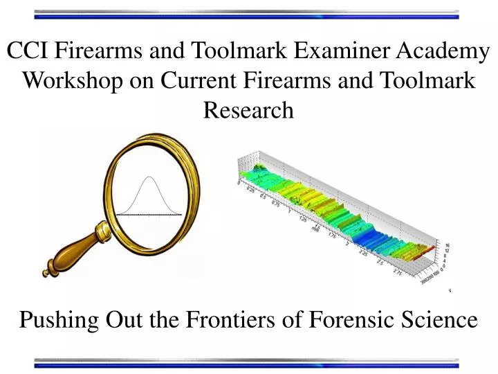
CCI Firearms and Toolmark Examiner Academy Workshop on Current Firearms and ToolmarkResearch Pushing Out the Frontiers of Forensic Science
Outline • Morning-ish • Introduction and the Daubert Standard • Confocal Microscopy • Focus Variation Microscopy • Interferometric Microscopy • Surface Data/Filtering
Outline • Afternoon-ish • Similarity scores and Cross-correlation functions • Known Match/Known Non-Match Similarity Score histograms. False Positives/False Negatives/Error Rates • Multivariate Discrimination of Toolmarks • Measures of “Match Quality” • Confidence • Posterior Error Rate/Random Match Probability • Lessons learned in conducting a successful research project
Introduction • DNA profiling the most successful application of statistics in forensic science. • Responsible for current interest in “raising standards” of other branches in forensics…?? • No protocols for the application of statistics to comparison of tool marks. • Our goal: application of objective, numerical computational pattern comparison to tool marks Caution: Statistics is not a panacea!!!!
The Daubert Standard • Daubert (1993)- Judges are the “gatekeepers” of scientific evidence. • Must determine if the science is reliable • Has empirical testing been done? • Falsifiability • Has the science been subject to peer review? • Are there known error rates? • Is there general acceptance? • Federal Government and 26(-ish) States are “Daubert States”
4 mm G. Petillo G. Petillo
5/8” Consecutively manufactured chisels Known Match Comparisons G. Petillo
5/8” Consecutively manufactured chisels Known NON Match Comparisons G. Petillo
5/8” Consecutively manufactured chisels 4 mm 4 mm 600 um
Confocal Microscope Marvin Minsky First confocal microscope
Source In focus light Illumination aperture Out of focus light Detector Confocal pinhole Objective lens Tool mark surface (profile of a striation pattern) Focal plane for objective Sample stage
Rastering pattern of laser confocal Nipkow disk sweeps many pinholes
Programmable array Illumination/Detection Get any illumination/detection pattern
Objective’s focal plane Scan stage in “z”-direction Sample stage
Detector Objective’s focal plane Scan stage in “z”-direction Sample stage
Detector Objective’s focal plane Scan stage in “z”-direction Sample stage
Detector Objective’s focal plane Scan stage in “z”-direction Sample stage
Detector Objective’s focal plane Scan stage in “z”-direction Sample stage
Detector Objective’s focal plane Scan stage in “z”-direction Sample stage
Detector Objective’s focal plane Scan stage in “z”-direction Sample stage
Detector Objective’s focal plane Scan stage in “z”-direction Sample stage
Detector Objective’s focal plane Scan stage in “z”-direction Sample stage
Detector Objective’s focal plane Scan stage in “z”-direction Sample stage
Detector Objective’s focal plane Scan stage in “z”-direction Sample stage
Detector Objective’s focal plane Scan stage in “z”-direction Sample stage
Detector Objective’s focal plane Scan stage in “z”-direction Sample stage
Detector Objective’s focal plane Scan stage in “z”-direction Sample stage
Detector Objective’s focal plane Scan stage in “z”-direction Sample stage
Detector Objective’s focal plane Scan stage in “z”-direction Sample stage
Detector Objective’s focal plane Scan stage in “z”-direction Sample stage
Detector Objective’s focal plane Scan stage in “z”-direction Sample stage
Detector Objective’s focal plane Scan stage in “z”-direction Sample stage
Detector Objective’s focal plane Scan stage in “z”-direction Sample stage
Detector Objective’s focal plane Scan stage in “z”-direction Sample stage
Detector Objective’s focal plane Scan stage in “z”-direction Sample stage
Detector Objective’s focal plane Scan stage in “z”-direction Sample stage
For Each Detector Pixel: Detector Point on surface corresponding to pixel’s is in maximum focus here Record the “axial response” as stage is moved along the z-direction
Increasing surface height All-in-Focus 2D Image Overlay confocal “z-stack”
Confocal Microscope Trivia • Use high NA objectives for best results • Small working distances • Flanks up to ~ 70o • Cost ~150K – 250K (FTI IBIS ~1M) • Get a vibration isolation table for your instrument ~7K • Set up in a (dry) basement if possible • Accuracy down to +/- 10 nm Optical slice thickness =
Confocal Microscope Trivia • Some manufactures: • Olympus • LEXT (Laser) • Zeiss • CSM (White Light) • LSM (Laser) • Nanofocus • msurf series (White Light) • Sensofar/Leica • Plu series/DCM (White Light)
Focus Variation Microscope “Low res” common Focus variation mic ~ +/- 1mm Scherer and Prantl
Detector In focus light Source Out of focus light Objective lens Tool mark surface (profile of a striation pattern) Focal plane for objective Sample stage
Cutaway Alicona, GMBH
Objective’s focal plane Scan stage in “z”-direction Sample stage
For Each Detector Pixel: Detector Point on surface corresponding to pixel is in maximum focus here Record the “axial response” as stage is moved along the z-direction
Focus Determination: Detector Pixel of interest Compute standard deviation (sd) of pixels grey values in the neighborhood A pixel in focus sits in a neighborhood with a large sd