Download
1 / 39
400 likes | 560 Views
Case 1 SF. 41 y/o woman with no PMHx p/w blurry vision and headache worsening x few weeks. No meds, no toxic habits. PE: Afeb VSS. Bilateral lid swelling. Neuro exam: MSE with mild cognitive slowing and diminished attention, otherwise normal. CN: diminished VA with disc swelling b/l.

E N D
Case 1 SF • 41 y/o woman with no PMHx p/w blurry vision and headache worsening x few weeks. No meds, no toxic habits. • PE: Afeb VSS. Bilateral lid swelling. • Neuro exam: MSE with mild cognitive slowing and diminished attention, otherwise normal. CN: diminished VA with disc swelling b/l. • Motor/sensory/cerebellar intact.
Other w/u • LP: elevated opening pressure, CSF lymphocytic pleocytosis (60-80), prt 80 • Serum ACE: ~65 • CSF ACE: ~20
Clinical course • Progressive cognitive decline, recurring bouts of aseptic meningitis with visual blurring partially responsive to IV steroids. • Poor compliance pulse Cytoxan • Later developed poor vision d/t glaucoma and b/l optic neuritis • Panhypopituitarism • Dementia
Case 2 RC • 44 y/o man with hx of CVA and behavioral problems, p/w AMS and difficulty walking. Prior CVA admitted at Montefiore 2005, p/w . W/u revealed basal ganglia and cerebellar calcification on CT scan, acute R midbrain/thalamic infarct on MRI. Cause of stroke in young uncertain, however pt found to be ANA+. • Meds:
PE: Afeb VSS Neuro: MSE: alert, Ox2, dim attention, STM 1/3. CN: dysarthric, otherwise intact Motor: increased tone in legs, full strength, mild incoordination on FTN, DTRs: hyperactive Gait: spastic/ataxic
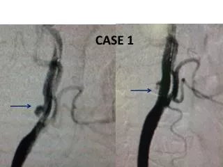
Other w/u • ESR 120 • Creatinine 2-3 (prior baseline 1-1.5) • ANA and dsDNA+ • Anti-cardiolipin Ab+ • MRI spinal cord: no significant abnl • Cerebral angiogram: possibly slight medium vessel irregularity c/w vasculopathy
Clinical course • IV steroids • IVIG • Mycophenolate • Warfarin • Worsening dementia and paraparesis • D/c to SNF
CNS Inflammatory Disease • Primary, recurrent demyelinating diseases: MS, Neuromyelitis Optica (NMO, Devic’s Dx) • Mono-phasic demyelinating diseases: Acute disseminated encephalomyelitis (ADEM), acute hemorrhagic leukoencephalitis (AHLE), transverse myelitis (TM), optic neuritis (ON); often these are para-infectious • CNS involvement with systemic (clinical or sub-clinical) auto-immune disease; includes primary and secondary CNS vasculitis • Paraneoplastic dx • Immune reconstitution inflammatory syndrome (IRIS) • CNS infections (discussed in other lecture)
Systemic inflammatory conditions with frequent neurological manifestations • SLE neuropsychiatric manifestations • Sjogren’s • Sarcoid • Anti-phospholipid Ab syndrome (1º or 2º) • Rheumatoid arthritis: PNS • Vasculitis: large or small vessel • Large: Giant cell arteritis: CN>CVA • Small: Wegener’s, polyarteritis nodosum: mononeuritis multiplex > CN >>CNS • Paraneoplastic syndromes: cerebellar dx, limbic encephalitis, PNS
Focal Clinical Presentation • Focal CNS deficit (brain or brainstem): hemiparesis, hemisensory loss, hemiataxia, diplopia, vertigo, dysarthria • Spinal cord syndrome: complete (motor/sensory/autonomic), anterior, posterior, Brown Sequard • Cranial nerve: optic neuritis, trigeminal neuralgia, facial paresis • Pseudo-peripheral: Lhermitte’s sign, paresthesias, pain • Focal cognitive deficit: aphasia, apraxia, neglect
Neuropyschiatric SLE: 19 syndromes describedJoseph (2007) Neurology
NPSLE • Neurological dx present in: ~50% (15-90%) • Presenting with neuro symptoms: 3-5% • NPSLE worsens prognosis • NPSLE can occur without systemic flare • Lab abnl: ESR elevated 50%, ANA+ 85%, dsDNA+ 72%, anti-phospholipid Ab 30%, complement low during flare 44%, ribosomal P Ab and C3A frequently elevated prior to/during flare. • APS associated with NPSLE, CVA, other focal dx
Neuro testing in NPSLE • CSF abnl: 20-40% (lymphocytic pleocytosis, elevated prt, OCB each present in ~20%). • EEG abnl: up to 80% abnl, mostly non-specific changes but some with epileptogenic focus. • EMG/NCS: high% abnl in symptomatic PNS dx
Neuroimaging • Brain MRI: abnl in 20-70%; most common findings are multifocal small white matter hyperintensities and atrophy; stroke in < 20%; lower % show basal ganglia calcification, reversible leukoencephalopathy syndrome (RPLS). • SPECT: detects multifocal or patchy/diffuse perfusion deficits in 50-90% • MR spectroscopy: abnl in ? 20-50%
EMT with SLE, APS, complicated migraine with aphasia and RHP
Neurosarcoidosis • Neurological manifestations in ~10% (~20% at autopsy). • Rarely presents with neurologic syndrome % • Very rarely limited to NS %
Laboratory findings in neurosarcoidosis • CXR abnl: ~40-50% (30-80% range) • Chest CT abnl: ~60-75% (? up to 90%) • Gallium/PET scan abnl: 25-80% • Serum ACE elevation: 25-75% • CSF prt elevation: 50% • CSF lymphocytic pleocytosis: 40% • CSF OCB: 20-40%
Neurosarcoid MRI abnl • Any abnl: up to 80% • Leptomeningeal or parenchymal enhancement: 25-50% • White matter lesions: 30-50%
Neurological manifestations of Sjogren’s syndrome • Common disorder, affecting ~2-3% of adults. • Neurological dx present in 5-60%. • CNS and PNS dx both common. • Neurological symptoms occur prior to diagnosis in 80-90% of patients. • Sicca symptoms present in <50% at presentation.
MRI, path, and sweat testing in Sjogren’s sensory neuropathy (Mori 2005)
Lab abnl in Neuro-Sjogren’s • SSA/SSB+: 45% • Schirmer’s test abnl: 90% • Salivary scintography abnl: 65% • Lip bx abnl: 95%
References • SLE: • Joseph (2008) JNNP • Sanna (2003) Lupus • Csepany et al (2003) J Neurol • Sjogren’s: • Mori (2005) Brain • Delalande (2004) Medicine • Soliotis (1999) Ann Rheum Dis • Sarcoid: • Joseph (2008) JNNP • Joseph (2007) Practical neurology • Spencer (2004) Sem Arthritis Rheum