Download
1 / 14
140 likes | 490 Views
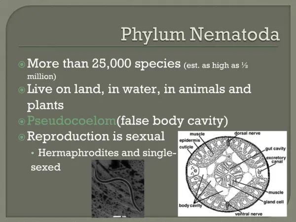
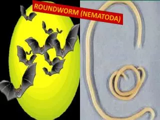
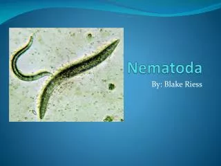
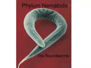
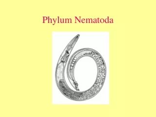
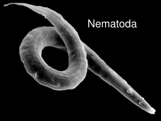
Class Nematoda. General characters. Cylindrical round worms with tapering ends. Separate sex, the female is usually larger than the male. No intermediate host. Infective stage: embryonated egg. Body is usually tapered to a pointed posterior end, and to a rounded anterior end.

E N D
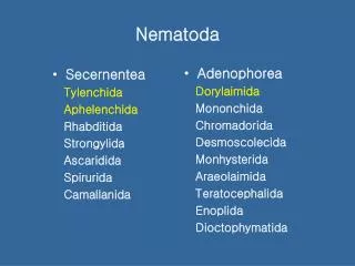
General characters • Cylindrical round worms with tapering ends. • Separate sex, the female is usually larger than the male. • No intermediate host. • Infective stage: embryonated egg. • Body is usually tapered to a pointed posterior end, and to a rounded anterior end. • They are classified into 2 main categories according to their primary location in the body: • Intestinal nematodes • Tissue nematodes (filariae)
Examples of Class Nematoda • Intestinal nematodes: Ascaris lumbricoides Enterobius vermicularis
1. Ascaris lumbricoides Egg :rounded with coarsly mamillated wall(H.P) Posterior end of male: curved with 2 spicules (L.P)
Ascaris lumbricoides cont. • Location of adult: Small intestine of man • Infective stage: Embryonated egg • Mode of transmission: Ingestion of food (green vegetables) contaminated with embryonated egg • Diagnosis: Eggs in stool • Disease: Ascariasis
2. Enterobius vermicularis (pin worm, oxyuris) Male (5mm): Posterior end is curved with one spicule (4X) Egg: Planoconvex or D shaped egg (H.P)
Enterobius vermicularis(pin worm, oxyuris) cont. • Location: Large intestine of man. • Infective stage: Embryonated egg. • Mode of transmission: Ingestion of food contaminated with embryonated egg or autoinfection via nails scratching the perianal region. • Diagnosis: Eggs in anal or perianal swab. collected using transparent adhesive tape. rarely in stool. • Disease: Enterobiasis.
Arthropods • Class Insecta: Flea, louse, bed bug • Class Arachnida:Hard tick • Class Crustacea:Cyclops • Question form:Name, medical importance
1. Flea • Composed of head with antenna, thorax carrying 3 pairs of legs, abdomen formed of 10 segments (4X) • Medical importance: • Vector of plague. • Intermediate host for Hymenolepis diminuta, Dipylidium caninum. • Transmit endemic typhus or murine typhus.
2. Bed bug Composed of head with antenna, thorax carrying 3 pairs of legs, abdomen formed of 8 segments,rounded end (4X) Medical importance: Insomnia, irritation
3. Louse • Composed of head with antenna, thorax carrying 3 pairs of legs, abdomen formed of 9 segments (incompletely segmented) carrying respiratory spiracles, bifid end (4X) • Medical importance: • Pediculosis • Could transmit trench fever and epidemic typhus
4. Hard tick • Composed of false head without antenna, thorax fused with head (cephalothorax), abdomen is not segmented, 4 pairs of legs ending by claws (4X) • Medical importance: • Mechanically: bite, bleeding • Systemically: tick paralysis • Could transmit rocky mountain spotted fever
5. Cyclops • Composed of cephalothorax carrying 1 median eye, 2 pairs of antennae, abdomen is segmented 4 or 5 segments, female carry 2 egg pouch, last segment carry legs (L.P) • Medical importance: • Intermediate host for: Diphylobothrium latum, Diphylobothrium mansoni, Dracunculus medinensis.
Microsporidia • Unicellular parasites • 1-40 μm: the smallest eukaryotes • Lack mitochondria and possess, mitosomes • Lack motile structures • Produce highly resistant spores • The spore is protected by a wall, consisting of 3 layers: • An outer electron-dense exospore • A median, wide and seemingly structureless endospore, containing chitin • A thin internal plasma membrane • A number of species may infect humans: these include Trachipleistophora hominis • Nowadays microsporidia are placed within the Fungi