Download
1 / 44
470 likes | 795 Views
emple Chemistry Department Philadelphia, PA www.chem.temple.edu. Biological Photochemistry: The fate of electronic excited states in proteins, DNA, and the role of quenching Robert J. Stanley DOE Workshop on Aqueous Scintillators January 19, 2010. Electronic excited states in Biology.

E N D
emple Chemistry Department Philadelphia, PA www.chem.temple.edu Biological Photochemistry: The fate of electronic excited states in proteins, DNA, and the role of quenching Robert J. Stanley DOE Workshop on Aqueous Scintillators January 19, 2010
Electronic excited states in Biology • Chemiluminescence • Bioluminescence – charge transfer? radicals? • Photoinduced electron transfer • Photosynthesis • DNA repair • Photochemistry • DNA damage • photosensors
5’ 3’ DNA…a polymer of nucleotides connected by phosphodiester linkages Nucleic acid bases A, T, C, & G Voet and Voet, Biochemistry, 2nd Ed. Wiley, New York, 1995
B-DNA is double-stranded (ds) DNA, forming the famous double helix (1954 - Watson, Crick, Franklin) Watson-Crick base pairing (complementarity)
DNA absorbs UV radiation * transition
Quenching of excited states can be desirous or devastating in living systems: DNA • UV light absorbed by DNA is rapidly transformed into heat • Conical intersections in the potential surfaces of excited and ground state nucleic acid bases leads to ultrafast degradation of light into heat (10-12 sec.) …GOOD! • Excited native DNA bases (Guanine, Adenine, Thymine, Cytosine) can be either excited state donors or acceptors • sequence dependent reaction • *G8-oxo-G • T-T T<>T pyrimidine dimerization • Cancer, apoptosis…BAD
H C 3 h CH 3 O O O O CH CH 3 3 HN NH HN NH O N O O O N N N T<>T or CPD T-T UV light damages DNA Bad photochemistry 2+2 photo-cycloaddition < 320 nm
If DNA damage is left unrepaired then mutations, cell death, and cancer can develop http://toms.gsfc.nasa.gov/ery_uv/euv.html
Bright Dark Bright or Dark Pathways involving energy transfer D*A D = G*, A*, C*, T* A = G, A, C, T hD DA* hA Triplet Energy Transfer Förster or Dexter Transfer (singlets) DA Fluorescence
“Structural” quenching pathways D*A Bright Dark DhotA hD Intramolecular vibrational relaxation Conical Intersection DA Fluorescence
Bright Dark Bright or Dark Pathways involving electron transfer D*A hD hEX? Photoinduced Electron Transfer (PET) Exciplex (EX) formation (charge transfer) DA Fluorescence
Enzymatic Repair of CPDs by DNA Photolyase uses blue light as an energy source (Good photochemistry) • Repair of the thymidines is direct: • T<>T T-T without modifying the DNA backbone • Wide spread: E. coli, Frogs, Rice, Kangaroos…Humans (no!) • Possible Applications: • Photosomes® (AGI Dermatics) • transgenic crops (wheat?) • Mees, A., et al (2004) Science 306, 1789-1793. Sancar, A. Structure and function of DNA photolyase. Biochemistry33, 2-9 (1994).
DNA Photolyase (PL) is aflavoprotein (Vitamin B2) that binds and repairs CPDs • PL functions efficiently with only FAD(required for repair and binding • PL binds the CPD with high affinity (no light required): • KA = 109 M-1 for dsDNA with CPD Park, H.-W., Kim, S.-T., Sancar, A., and Deisenhofer, J. (1995) Science268, 1866-72.
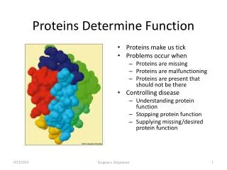
FADH— — Flavin Structure and Oxidation States • Flavins can transfer 1 or 2 electrons (unlike nicotinamide) and are used in a large number of redox reactions in the cell • Surprisingly, flavins are a major biological chromophore (DNA repair, circadian rhythms, phototropism, etc.) Biochemistry 2nd Ed., Voet and Voet, J. Wiley & Sons
A large separation between the FADH- and the CPD (~16 Å) would give a slow electron transfer rate (keT, from Marcus theory) Orbital overlap x Driving force • Slow electron transfer would compete poorly with 1FADH— deactivation (about 5 ns) • but repair > 0.7! Photolyase functions by Photoinduced Electron Transfer from the FAD to the CPD There’s a cavity in the protein FAD
What happens to substrate conformation upon binding to Photolyase? Minor disruption AA Photolyase Moderate disruption Base Flipping T<>T Severe disruption
3’ 5’ 3’ 5’ 5’ 3’ 5’ 3’ 3’ 5’ 3’ 5’ 5’ 3’ 5’ 3’ Fluorescent reporter approach to probing double helical structure 5’-probe approach: Base Flipping 3’-probe approach: Base Flipping The fluorescence quantum yield of the reporter decreases when base stacked…but why?
6MAP is an attractive new fluorescent adenosine analogue 4-amino-6-methyl-8-(2-deoxy--D-ribofuranosyl)-7(8H)-pteridone Properties:1 fl = 0.2 ex = 330 nm ( ~ 8,500 M-1cm-1) em= 430 nm (large Stokes shift) 1Hawkins, et al, “Synthesis and Fluorescence Characterization of Pteridine Adenosine Nucleoside Analogs for DNA Incorporation.” Anal. Biochem.298, 231-240 (2001). K. Yang, S. Matsika, and R.J. Stanley, Biochemistry 2007
Base flipping of the CPD monitored by 6MAP +PL -PL 5’-GCAAGTTGGAG-3’ 3’-CGTTCAFCCTC-5’ +PL 5’-GCAAGTTGGAG-3’ 3’-CGTTCFACCTC-5’ -PL Why is the intensity pattern sequence-dependent?
Photolyase These data are consistent with disruption of base stacking due to base flipping of the CPD by Photolyase ? Mees et al, Science v. 306, 1789-1793 (2004)
Is the fluorescence quantum yield modulation of 6MAP due to PET? Stern-Volmer quenching of 6MAP by G,A,C, and T: what is the rate of quenching, kq? What are the redox potentials? Cyclic voltammetry of 6MAP in aprotic organic solvents submitted to Biochemistry
The quenching of 6MAP* proceeds through nucleobase oxidation: 6MAP*:NMP6MAP¯:NMP+ (Scandola-Balzani relation) submitted to Biochemistry
What’s the mechanism for base analog quenching?Pathways for energy transduction in a model FBA oligo Absorption Stark spectra of ssDNA with 2AP (), a hexamer with 2AP () , and a mix of the individual bases (). Stark and MRCI calculations (Matsika) Stark absorption and emission spectra of 6-MI (), a guanine analog, compared with their absorption and emission spectra ().
Another possibility: 6MAP emission overlaps the absorption of the FAD: FRET from 6MAP*FAD? Yang et al, JPC B (2007)
Fluorescence Energy Transfer Efficiency R0 the Förster distance where ET = 0.5 rDA the distance between a donor (fluorescent analogue) and an acceptor (FAD in photolyase)
R0 (Å) = The Förster distance 2 : the orientation factor; n : the refractive index of the medium; D : the fluorescence quantum yield of the donor; J : the overlap integral.
The Overlap Integral FD(): the fluorescence intensity of the donor as a function of wavelength. εA(): the molar extinction coefficient of the acceptor at that wavelength;
The Orientation Factor mD mA rDA θT: mD, mA θD: mD , rDA θA: mA, rDA
The transition dipole moment direction 6MAP was calculated from TD-DFT Yang et al, JPC B (2007)
Orientation factors and ETbetween Probes and FADox From the crystal structure, lit. and TDDFT calcs experiment crystal structure Yang et al, JPC B (2007)
FRET efficiency vs. orientation Yang et al, JPC B (2007)
NO FRET! • The FAD is quenched 100x in the protein (acceptor is dark) • A work-around : time-resolved FRET? • Quenching mechanism is different for the two probes • photoinduced electron transfer vs. ultrafast internal conversion? • Does FAD* undergo PET to tryptophan??? Yang et al, JPC B (2007)
1PLred : T<>T 2 eT PLsq• : T<>T • kic, krad 3 1 PLsq• : T|_|T • 2 4 krec PLsq• : T-T • kbeT 5 PLred : T-T kdiss 6 PLred + T-T 1 PLred : T<>T 7 Can we identify the kinetics and mechanism of repair? Two color pump probe femtosecond spectroscopy: • What is the electron transfer lifetime (eT)? • Does repair proceed by a concerted or sequential mechanism? c MacFarlane and Stanley (2003) Biochemistry42, 8558-8568
F1 M9 Laser control Mode and wavelength monitor W2 M10 M8 M11 M13 P1 Delay stage controller W1 M2 W3 L3 Ti:sapphire B1 M12 F2 L4 ISO M6 M M3 M7 L1 CW Nd:YAG M1 L2 L5 Monochromator Ti:Sapphire amplifier L6 M4 CCD M15 M5 L7 L8 YLF laser M14 Chopper Controller Synchronization Delay Generator Transient absorption measurement layout BBO CaF2 Sample
PET to the CPD substrate quenches the FADH excited state in ~ 30 ps MacFarlane and Stanley (2003) Biochemistry42, 8558-8568
1PLred : T<>T 2 keT krad PLsq• : T<>T • 3 krepair PLsq• : T-T • 4 krec 1 PLred : T<>T or T-T What’s are the intermediates? A unidirectional sequential model: • A(,t) = ci(t)i() = C(E - 0) • where Ei() = True spectra of the intermediates • 0() = Ground state absorption spectrum • Construct C(t) = C0eKt (from the K matrix) • Calculate Ei () = C-1A(,t) • Minimize {A(,t) – C(E- 0)} using K matrix
The broadband transient absorption data: Pl-red+(TTT<>TT) Pl-red+(TTTTT)
PLSQ Spectrotemporal intermediates in the repair reaction: E spectra 1PLred : T<>T 53 ps 2 PLsq• : T<>T • 3 540 ps 620 ps PLsq• : T-T • 4 2753 ps 1 PLred : T<>T or T-T • Fitting the data does not rule out a sequential bond breaking mechanism... • More complicated kinetics cannot be ruled out!
Bright Dark Bright or Dark D*A h Photoinduced Electron Transfer (PET) DA Fluorescence In conclusion…Quenching is a simple term for many possible mechanisms to shunt electronic energy in excited molecules A battery of approaches need to be used to explore all possible pathways
The Charge Separation Investigation Team • Madhavan Narayanan • Ultrafast spectroscopy • Protein Chemistry • Dr. Zhanjia Hou • Ultrafast spectroscopy • Single molecule spectroscopy • Goutham Kodali • Stark spectroscopy • Computational chemistry • “Vector dude” • Dr. Alex MacFarlane IV • Ultrafast spectroscopy • Electric field effects • Salim Siddiqui, M.D., Ph.D. • Stark spectroscopy • Computational chemistry
Gone, but not forgotten.. The Group Funding NSF Molecular Biosciences, REU Petroleum Research Fund Collaborators Prof. Aziz Sancar (UNC) Mary Hawkins (NIH) Prof. Spiridoula Matsika
2.4Å 1.9Å Watson-Crick base pairing is distorted Base stacking is weakened A closer look at the damage… 5’-GCTTAATTCG-3’ 3’-CGAATTAAGC-5’ A A 5’ 3’ Crystal structure: Park et al, PNAS99, 15965-15970 (2002).
DNA Photolyase (PL) binds its CPD substrate by base flipping CPD Flavin Adenine Dinucleotide • Mees, A., et al (2004) Science 306, 1789-1793.
Spectral overlaps of probes and FAD S0S2 S0S1 Does FRET explain the intensity pattern difference?