Download
1 / 24
350 likes | 1.48k Views
Thromboembolic disease in pregnancy. Dr Hashmi hajrasi Consultant in OBS & GYN MBBCh , DGO, MRCOG, D’MAS. Lecture objectives. By the end of this lecture, student is expected to Know T he implication of thrombo -embolic disease(TED) on pregnant women

E N D
Thromboembolic disease in pregnancy Dr Hashmihajrasi Consultant in OBS & GYN MBBCh, DGO, MRCOG, D’MAS
Lecture objectives By the end of this lecture, student is expected to Know • The implication of thrombo-embolic disease(TED) on pregnant women • Why pregnancy is associated with increased tendency for clotting • Risk factors for TED • Clinical Symptoms & signs of DVT and diagnostic difficulties
Types of DVT • Diagnostic tests • Treatment of acute phase DVT & subsequent management • Clinical presentation of pulmonary embolism, symptoms & signs and confirmatory lab tests • Management of PE • conclusion
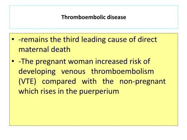
Introduction • Venous TED is one of the major causes of direct maternal deaths. Those who survive suffer significant morbidity • Pregnancy is associated with 5-6 fold increased risk of TED than outside pregnancy. The true incidence is unknown but rated between 0.3 – 1.2% of all pregnancies • In 80% of cases it occurs post-nataly usually during the first 2 weeks and in 80% of cases it is left-sided
Why pregnancy is associated with increased tendency for clotting ? Virshow’s triad • Venous stasis • Increased production of clotting factors • Increased tendency for platelet aggregation
Risk factors for TED • Age over 35 yrs • Multi parity ( ≥ 4) • Obesity ( over 80 kg) • PET • Immobility • Personal or family H/O TED • Thrombophilia • Operative delivery (em. C/S > elective )
Gross varicose veins • Blood group other than O
Types of venous thrombosis • Superficial thrombo phlebitis • Calf (below knee)deep vein thrombosis • Proximal or (ilio-femoral) deep venous thrombosis
Diagnosis • Because of future implications ,risks , treatment costs and inconvenience, every effort should be made to have an objective diagnosis. However, • Clinical diagnosis is inaccurate in over 60% of cases of TED
Superficial thrombophlebitis • The condition is misnamed. It is not infective. the redness surrounding the affected vein is a reaction to clot • It is the commonest form of venous thrombosis in pregnancy & puerperium. It occurs in about 1% of patients and nearly always arise in existing varicose veins • The diagnosis is clinically obvious (tenderness, erythema, palpable cord-like veins
Treatment is usually symptomatic with compression bandage, leg elevation and to encourage mobility • In some pt’s DVT need to be excluded as it may co-exist with it . evenmore extension to involve deep veins rarely occurs
Calf deep venous thrombosis (CVT) • The most common clinical features are pain, local tenderness, swelling, change in skin colour and temperature • Most of CVT resolve spontaneously (75-80%) and run a benign course except when the thrombus spreads up to involve the proximal deep veins (20-25%) in which case there is 50% risk of pulmonary embolism
Proximal DVT • It occurs more commonly than CVT and over 80% is left-sided • Symptoms are more dramatic with pain and swelling involving the entire limb • If the arterial supply is unimpaired, the leg appears swollen, blue & warm. On the other hand if arterial spasm occurs secondary to irritation from the nearby clotted vein, the leg becomes swollen, painful, white & cold
Pulmonary embolism (PE) • A high index of suspicion is always needed for the diagnosis of PE especially in patients with DVT or risk factors for VTE • The maternal mortality rate from untreated PE is 13% with the majority within 1 hr of the event • With early diagnosis & treatment, the survival rate is between 92-95%
The common symptoms & signs of PTE • Tachypnoea • dyspnoea • Haemoptysis • Pleuritic chest pain • tachycardia • Cyanosis • Pyrexia • Syncope or varying degree of shock These S &S are non-specific and in most cases there is no prior clinical evidence of DVT
Investigations for suspected PTE • Chest X- ray • ECG • Blood gases • Compression duplex Doppler to exclude DVT • D-dimer is used as a screening test for VTE with high negative predictive value. A low level suggests absence of VTE and further objective tests are not needed
Ventilation-perfusion isotope lung scan (V/Q) • Helical or spiral CT scan is regarded superior to V/Q scan • computed tomograhy pulmonary angiogram (CTPA) if clinical suspicion is high but other tests are inconclusive
Treatment of acute phase TED • Standard heparin IV or the more preferred LMWH S.C should be started once the diagnosis is clinically suspected until excluded by objective testing • Treatment aims at achieving APTT 2-2.5 the control for 5-7 days then continue with prophylactic dose generally for 6-12 weeks post-nataly
Heparin is the anticoagulant of choice in pregnancy. It does not cross the placenta and in overdose action can be reversed by protamin sulphate • Osteoporosis & thrombocytopenia are complications of prolonged heparin treatment. Therefore platelet count should be monitored regularly
Legs should be elevated & graduated elastic compression stocking should be worn to reduce oedema • In DVT, calf circumference should measured daily to help monitoring the response to treatment • Massive PE requires ICU & multi disciplinary team approach • Recurrent PE may require inferior vena cava filter
Thrombolytic therapy in PE should only be given with haematologist agreement • Thoracotomy with embolectoy may be life saving • Heparin thrombo -prophylaxis has to be considered in the subsequent pregnancies or if additional risk factors appear
Oral anticoagulants • Cross the placenta and are potentially teratogenic at any stage of pregnancy • Complications of warfarin includes, nasal hypoplasia, depressed nasal bridge, irregular bone growth & intracranial fetal haemorrhage • However , they can be given after delivery and are safe for lactation
conclusion • Thrombo-embolism is amajor cause of maternal mortality &morbidity worldwide • Clinical diagnosis is unreliable but once strongly suspected, treatment should be started until objectively excluded • Dupplex Doppler, x-ray venogram & V/Q scan are the main diagnostic tools
During pregnancy, LMWH is the preferred anticoagulant as it is more effective and safer than standard heparin • Oral anticoagulants should not be given at any stage during pregnancy but they are safe & may be more convenient after delivery • High clinical suspicion with early full anticoagulation and objective diagnosis are the best ways to minimize maternal M&M and avoiding risks of the unnecessary treatment