Download
1 / 85
850 likes | 859 Views
SEMINAR ON LUPUS ERYTHEMATOSUS. The Clinical spectrum of Lupus erythematosus is defined by cutaneous LE limited to the skin at the one extreme and by multisystem disease with diffuse proliferative glomerulonephritis at the other. Cazenave introduced the name LE in 1851.

E N D
SEMINAR ON LUPUS ERYTHEMATOSUS
The Clinical spectrum of Lupus erythematosus is defined by cutaneous LE limited to the skin at the one extreme and by multisystem disease with diffuse proliferative glomerulonephritis at the other.
Cazenave introduced the name LE in 1851. Kaposi was first to recognise the systemic components of LE
Epidemioloigy -Skin disease is thesecond most frequent clinical manifestation of LE after joint inflammation. -All races are affected -ACLE like SLE is more common in women than men(8:1) -SLE is 7fold more common than DLE.
SCLE is primarily a disease of white females, with the mean age of onset in the fifth decade. DLE can occur in infants and the elderly, it is most common in individuals between 20 and 40 years of age. DLE has a female:male ratio of 3:2 to 3:1, more prevalent in blacks. 90% of SLE patients are women of Child bearing age group, highest prevalence is seen in African and Americans.
Revised ARA Criteria • Malar Rash • Discoid Rash • Photo sensitivity • Oral ulcer • Arthritis • Serositis • Renal disorder • Neurologic disorder • Hematologic disorder • Immunologic disorder • Anti Nuclear antibodies
Etiopathogenesis • 4 theoretical phases 1. Susceptibility phase: involves the inheritance of gene that confer predisposition to injury. HLA class II antigen are more clossely assosiated with sp.autoantibody. Genes for complement proteins & TNF, genes mediating apoptosis, Gene involved in T-cell signalling process and genes for Clearance of immune complex are implicated.
2. Induction phase: involves the initiation of autoimmunity characterised by the appearance of auto reactive T-Cell that exhibit the loss of self tolerance. Envrionmental factors play an important role.
3. Expansion stage: involves progressive augmentation of the autoimmune response and has feature of an antigen driven response. Serological detectable auto antibodies are produced by the population of clonally expanded B-cell.
Three major target of antibodies: • Nucleosome- anti DNA & anti histone antibody • Splicesome – anti Sm & anti RNP As • Ro & La molecules – Anti Ro & La.
4. Immunological injury: This phase can be attributed to the action of auto antibodies and the immune complex that form, which causes tissue damage by direct cell death, cellular activation, opsonization and blocking of the target molecule functions.
Role of genetics in LE specific skin disease: SLE- HLA DR2, DR-3, B8 SCLE – HLA B8, DR-3 (annular), DR-2 (papulo squamous) Genetic deficiency of complement components including C2-5 & C1 esterase inhibitor deficiency SCLE & DLE DLE- HLA B7, B8, DR2, DR3 & DQA 0102. Cutaneous LE IL1 receptor antagonist & TNFα gene polymorphism
Role of UV light in LE specific skin disease: UV Light: Most important environmental factor in the induction phase. Uncover an already established auto immune state by triggering auto Ag release from injured keratinocytes. Have early critical role in the induction phase by altering cellular DNA.
Affect localization of preexisting autoantibody containing immune complex by altering endothelial permeability UV induced keratinocyte apoptosis-responsible for aberrant pattern of cell surface auto Ag expression. Exaggerated release of immune mediators like IL1, TNFα PG-E, protease, O2 free radicals & histamines
Role of Tobacco exposure: • Smoking- ↑ risk of developing SLE. • Related to lupogenic aromatic amines contained in tobacco smoke. Other Factors: Heavy metals, dietary supplements of alfalfa,silica,infection,stress,hormonal factors Late menarche is associated with increased risk of SLE Infection of all types of viruses are known to exacerbate SLE.
Role of Drugs: • Allopurinol, Atenolol, Captopril, carbamazepine, chlorpromazine, gold, hydralazine, hydrochlorothiazide, INH, Lithium, methyldopa, minocycline, nitrofurantoin, OCP, penicillamine, Phenyl butazone, phenytoin, phenothiazine, procainamide, propranalol, sulfonamides, tetracycline & Valproate.
Drug induced LE: 82% have anti histone Ab • ↑ incidence in HLA DR-4, more common in females(4:1). • > in slow acetylators. • Similar manifestation to SLE, without cutaneous involvement • Hydralazine: inhibits binding of C4
Drug induced SCLE-skin is often involved • DRUGS Hydrochlorothiazide, CCB, ACE, Terbinafine, piroxicam, d-penicillamine, sulfonylureas, oxprenalol, parental gold, Spironolactone, IFNβ & Ranitidine.
LE specific skin lesions • Chronic Cutaneous LE(CCLE): • 1.DLE. • Skin lesions: discrete erythematous plaques, well defined adherent scale that extend to dilated hair follicles. Heal with atrophy, telangiectasia in the centre.
Scalp involvement- alopecia (follicular distruction) • Mucous membrane involvement in 15%. • Disseminated DLE: lesions occur both above and below neck. • 1.3- 6.5% localised & 22% disseminated DLE cases develop SLE. • 1/4th patients with SLE develop DLE lesions in some point in the course of their illness
Clinical features that suggest SLE in patients with DLE skin lesions: • History: Unexplained fever, Extreme fatigue, joint pain, raynauds phenomenon, pleuritic chest pain, photosensitivity, LE non specific but disease related skin lesions.
Physical findings: Diffuse hair loss, lymphadenopathy, SCLE/ACLE skin lesions. • Lab anomalies: unexplained anemia, Leucopenia, ↑ESR, positive ANA, hyper gamma globulinemia, Immune deposit at DEJ of clinically normal skin.
Histopathology: • Basal/germinal cells of epidermis is the principal site in injury. • Prominent hyperkeratosis, well developed follicular plugging, nucleated layer of epidermis may be thinned out. • Loss of normal organization of basal cells; • Edema with vacuole formation within basal cells.
Partial obliteration of DEJ by a mononuclear cell infiltrate. • Accumulation of melanin in dermal macrophages. • Liquifactive degeneratin of the basal cell layer. • Mononuclear infiltrate in dermis composed predominant of T-lymphocyte, localized to periappendegeal and perivascular areas.
Immunopathology of DLE • Thick band of Ig along DEJ in lesional skin. • Deposits are heavy and contain several Ig classes. • Homogeneous, thready or granular pattern • Do not occur in uninvolved skin. • In scarring alopecia, deposits occur around hair follicles.
Differential Diagnosis: • Plaque type of PLE, Granuloma Facie, Sarcoidosis, Jesners lymphocytic infiltrate, pseudolymphoma, lymphocytoma cutis, lupus vulgaris, tertiary syphilis • Hypertrophic LE: Keratoacanthoma, SCC, Prurigo nodularis, hypertrophic LP.
Laboratory Findings • Low titre ANA in 30%. • 5% will have positive ANA at significant titre. • Small percentage will have false +VDRL, RA factor, slight decrease in serum complement, moderate increase in Ig, modest leucopenia, and even antiphospholid Ab.
CCLE- OTHER FORMS LE panniculitis/Lupus profundus • Deep dermal and subcutaneous inflammatory involvement. • Deep, firm, nodules often without surface changes. • Histopathology: Insignificant epidermal involvement, perivascular lymphocytic involvement in deep dermis and SC fat, vessel wall thickening, prominent fibrinoid degeneration of collagen and calcification..
2.Hypertrophic or Verrucous DLE: • Extensor aspect of arm, upperback and face. • Overlapping features of hypertrophic LP and LE have been described. 3. MUCOSAL LE: Oral, nasal, conjunctival and genital. Oral mucosal LE can degenerate to SCC similar to long standing cutaneous DLE lesions.
4.Chilblain/perniotic LE: Precipitated by cold, damp climate. Associated with Ro, SSA Ab. 20%SLE. 5.Tumid LE:Succulent, edematous urticaria appear in plaques with little surface changes. • On histology, typical dermal findings of DLE are seen and characteristics epidermal histological changes of LE specific skin disease are minimally expressed.
Nail changes: Red blue colour of the nail plate, longtitudional striae,crumbling of nails, subungual hyperkeratosis. Rowell syndrome: Cutaneous LE occur with lesions resembling EMF on face, neck, chest and mouth.
Subacute Cutaneous LE • Antibodies to the Ro, SSA antigens are closely associated. • Comprise approximately 10% of patients with LE. • 2 types: papulosquamous(2/3), annular polycyclic(1/3) • Predominantly adults. • Usually occur above the waist, around the neck, on the trunk, and on outer aspect of the arm. • 50% fulfill ARA criteria for SLE. • Fever, Malaise and CNS involvement occur, but renal disease is mild and infrequent.
SCLE VS DLE • SCLE: less scarring less prominent scaling and follicular plugging, less persistent, more wide spreading distribution, less discrete. • More promninent pigmentary changes, but not associated with dermal atrophy. • Photosensitivity is very common.
LE lesions of hard palate in 40%. • Diffuse, nonscarring alopecia. • Livedo reticularis and periungual telangiectasia more in SCLE. • Almost all patients have mild systemic complaints: joint pain, unexplained fever, malaise. • 20% will have DLE/ACLE lesions.
Pathology • Similar to DLE, however follicular plugging, hyperkeratosis, and density and depth of cellular infiltrate are considerably less. • The mononuclear cell infiltrate is usually restricted to the perivascualr and periappendigeal areas of upper third of dermis.
The diagnostic findings are found along the basal layer of the epidermis, as seen in all forms of LE specific skin disease. • Pilosebaceous atrophy is the only significant preidctor of DLE vs SLE.
Immunopathology • In contrast to the frequent finding of Ig deposits of DLE, 40-50% of SCLE lesions do not have immune deposits at DEJ. • IgG is deposited in a dust like pattern over the nucleus and cytoplasm of epidermal keratinocyte and cells in upper dermis.
D/D: Papulosquamous SCLE: Photo sensitive variant of psoriasis. Annular SCLE: EMF, EAC, EGR, GA. Seborrhoeic dermatitis, PMLE, Dermatophyte infection, Nummular Eczema, CD, Dermatomyositis, PRP, Disseminated superficial actinic porokeratosis, Cutaneous lymphoma/Mycosis fungoidis.
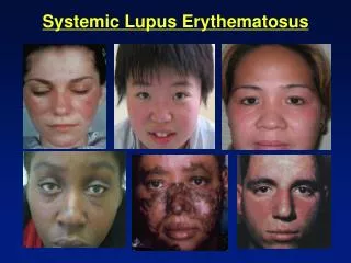
Acute Cutaneous LE(ACLE) • Localized ACLE: Classic butterfly rash or malar rash of SLE. • Confluent symmetric erythema and edema is centred over the malar eminence and bridge of the nose, Characteristically sparing the nasolabial fold. • Forehead, chin and V area of the neck can be involved and severe facial swelling can occur.
Generalized ACLE: Wide spread morbilliform or exanthematous eruptions often focussed on extensor aspect of arms and hands characteristically sparing the knuckles. An extremely acute form of ACLE, simulating TEN can occur.
ACLE lesions are abrupt in onset, last for hours to days, and frequently coincide with systemic disease exacerbation. • Post inflammatory hyper pigmentation is common, no scarring. • ACLE occasionally occur in conjunction to SCLE, but unusual with DLE.
Histopathologically: • Early phase unimpressive and non diagnostic. Careful examination will revive the basal layer changes characteristic of cutaneous LE. Dermal cellular infiltratives is usually sparse and the most prominent change being upper dermal edema and focal areas of basal area injury. Upper dermis: pronounced mucinosis. Epidermal necrosis can occur.
DIF: • Immune deposits at DEJ in skin lesions(90%) and uninvolved skin(60%) D/D: • Localized ACLE-Rosacea, Dermatomyositis, Seborrhoeic dermatitis, PMLE, Photo allergic CD. • Generalized ACLE-Drug hypersensitivity,Viral exanthem.
LE Nonspecific skin lesions: • Their presence indicates skin disease. • Vascular lesions- most common; telengiectasia(Linear nail fold capillary dilatation) • Deepset papular, palmar and distal telengiectasic lesions are characteristic of LE • Dermal vasculitis with ischaemic infarcts. • Recurrent superficial and deep thrombophlebitis may be an early sign. • 20% can have raynauds phenomenon( associated with good prognosis).
5% will have rheumatoid nodules. • A deforming non erosive arthritis(Jaccoud’s arthritis may be seen in these patients. They have high frequency of endocarditis. • Hair loss:- • CCLE-scarring alopecia.
SLE-frontal alopecia with increased fragility producing short, broken of hairs(lupus hairs) • Mucous membrane lesions usually occurs during acute flares. • Diffuse cutaneous hyper pigmentation may be seen. • Urticarial like eruptions may be seen. • Others: EMF, papulonodular mucinosis, PCT.
A. Bullous Lesions in LE: • ACLE- TEN like ACLE 2. SCLE: TEN like SCLE Vesiculo bullous annular SCLE 3. CCLE: Bullous DLE
B. LE Non specific vesiculobullous LE: 1. Bullous SLE 2. Vesiculo bullous disorder anecdotaly occur in LE BP, DH, PE, PCT