Download
1 / 23
300 likes | 1.02k Views
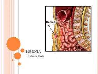
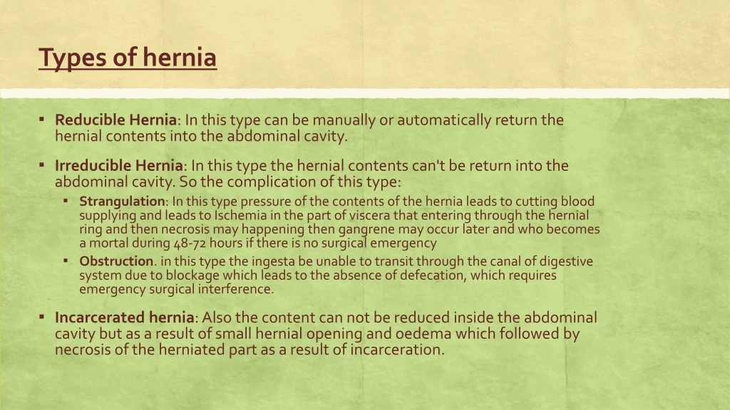
Types of hernia. Reducible Hernia : In this type can be manually or automatically return the hernial contents into the abdominal cavity. Irreducible Hernia : In this type the hernial contents can't be return into the abdominal cavity. So the complication of this type:

E N D
Types of hernia • Reducible Hernia: In this type can be manually or automatically return the hernial contents into the abdominal cavity. • Irreducible Hernia: In this type the hernial contents can't be return into the abdominal cavity. So the complication of this type: • Strangulation: In this type pressure of the contents of the hernia leads to cutting blood supplying and leads to Ischemia in the part of viscera that entering through the hernial ring and then necrosis may happening then gangrene may occur later and who becomes a mortal during 48-72 hours if there is no surgical emergency • Obstruction. in this type the ingesta be unable to transit through the canal of digestive system due to blockage which leads to the absence of defecation, which requires emergency surgical interference. • Incarcerated hernia: Also the content can not be reduced inside the abdominal cavity but as a result of small hernial opening and oedema which followed by necrosis of the herniated part as a result of incarceration.
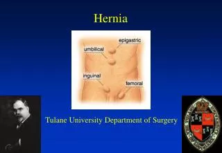
According to the topographical anatomy of the hernia • Umbilical hernia. • Abdominal hernia. • Inguinal hernia. • Scrotal hernia. • Perineal hernia. • Diaphragmatic hernia.
Umbilical hernia • It is secondary to failure of the normal closure of the umbilical ring and result in protrusion of abdominal contents into the overlying subcutis. Size varies depending on the extent of the umbilical defect and the amount of abdominal contents contained within it. • The aetiology in both large and small animals is likely to have a genetic component; however, excess traction on an oversized fetus or cutting the umbilical cord too close to the abdominal wall are other possible causes. • The umbilicus in new-born calves consists of the urachus (a tube that attaches the fetal bladder to the placental sac) and the remnants of the umbilical vessels that transport blood between the fetus and its mother. Normally, just after birth these structures shrink until only tiny remnants remain within the abdomen. If the area in the body wall through which these structures passed remains open, abdominal contents can protrude through the defect resulting in an umbilical hernia.
Causes of hernia specifically in pigs: • Environmental factors can increase the incidence of umbilical hernias so if there is a problem (more than 2% of pigs) consider the following: • Are prostaglandins used to synchronise farrowings. If so check that piglets are not being pulled away from the sow at farrowing and the cord stretched excessively. • Is navel bleeding occurring on the farm? Are naval clips being used to prevent bleeding? If so make sure they are not placed close up to the skin otherwise the tissues will be damaged and weakened. • Identify the precise time when the ruptures appear. Do these coincide with a change of housing? • In housing where the pigs pass through a small hole to the dunging area sudden severe abdominal pressure may cause ruptures. • Are stocking densities high and causing increased abdominal pressure? • In cold weather do the pigs huddle thereby increasing abdominal pressure? • Check records to see if any particular boar is implicated. • If the rupture is large and the pig is on a concrete floor or slats it should be moved to a soft bedded area so that the overlying skin does not become sore and ulcerated. • Examine navels at births and two days later to see if there are any abnormalities.
Umbilical hernias can be divided into: • Uncomplicated hernias • those whose contents can be easily pushed back into the abdomen. • Strangulated hernias • when contents cannot be easily pushed back into the abdomen, usually firm and may be painful to touch. The animal may show signs of colic and/or bloat and may die from the condition. • Umbilical hernias • with an associated abscess comprise two distinct parts: the abscess component is a firm lump adhered to the skin and the hernia is a softer swelling above this. Hernias with associated abscesses usually begin as an infected umbilicus alone (navel ill)
Multiple births and shortened gestation lengths are two important risk factors for congenital umbilical hernias in calves. • Hernia size varies depending on the extent of the umbilical defect and the amount of abdominal contents contained within it. Umbilical hernias are the most common birth defects in calves, especially in Holstein-Friesians. • .Most umbilical hernias are repaired with minimal surgical intervention.
Scoring umbilical hernia in pig Score 0 Score 1 Score 5 • Umbilical scores were assigned on a scale from 0 to 5. • Zero = no signs of swelling/ hernia • 3 = no hernia, no scab, but hard to the touch • 5 = hernia present.
Abdominal hernia • Is a term used to describe hernia that occurs in the abdominal wall other than those occurs through a natural orifice. It usually results from rupture of the abdominal muscles and aponeurosis. • This form is common in horses, cattle and swine (pig #2 in the surgery) • The most probable cause is a trauma as a kick from a neighbouring animal. A horn trauma usually results in subcutaneous rupture of the abdominal muscles. • Abnormal contraction of the abdominal wall as in cases of advanced pregnancy.
Abdominal hernias are seen high or low in the flank, along the costal arch or in the ventral abdominal wall in front or behind the umbilicus. • The hernial contents are either small intestine, large intestine, omentum, uterus, abomasum or urinary bladder. • In the beginning of the condition there is an inflammatory swelling with oedema that it is rather difficult to diagnose the case. • Later on the characteristic symptoms of a hernia well developed.
Scrotal hernia • Is the displacement of the part of the intestine or omentum from the abdominal cavity through the internal inguinal ring, inguinal canal, external inguinal ring and stay in the vaginal cavity inside the scrotum beside the testicle. • Mostly the scrotal hernias are inherited. • The same causes which cause inguinal hernia may cause scrotal hernia directly or the scrotal hernia follow the inguinal hernia. • Upon physical examinations there is a unilateral enlargement of the scrotal sac in the rear end of the hindlimb. Hernial content is soft and painful. The hernial content is usually reducible. Testicle atrophied. Inguinal ring can be easily felt and you can introduce 2 - 3 fingers inside it.
Inguinal hernia • In male pigs are common, and they usually extend into the scrotum. • Suspending the piglet by the forelegs and gently shaking, which generally causes even a small hernial bulge to become visible can confirm the diagnosis. • In female pigs, this defect is invariably accompanied by arrested genital development; such animals are sterile, and surgery is indicated only when the size of the defect is a threat to the growth of the pig to market weight. https://www.youtube.com/watch?v=tqBgyORH1MM
Congenital hernias are rarely seen in male cattle. It is evident in early calf-hood and are occasionally encountered as intestinal evisceration following castration. • Acquired inguinal hernias may be of traumatic origin or may result from management practices in the development of young bulls. Traumatic inguinal hernias have occurred in bulls that have become entangled when attempting to jump fences. Perhaps in these cases increased intra-abdominal pressure allows the intestines to perforate the peritoneum near the vaginal ring and descend through the inguinal canal alongside the spermatic cord. The intestine frequently become strangulated. • Surgical correction to preserve the breeding potential of the bull, when done, is not always successful.
Perineal hernia • It is a lateral protrusion of a peritoneally lined hernial sac between the levator ani and either the external anal sphincter muscle or the coccygeus muscle. • In sows the whole of the perineal region can present in collapse. The rectum and vagina may prolapse into the hernia. The hernia can be very large. There is no economic treatment. If the sow is close to farrowing keeping until farrowing may be an option, however, manual removal of piglets are likely to be required. Provide the sow with a bran diet or add liquid paraffin from time to time to help with the passage of faeces until slaughter
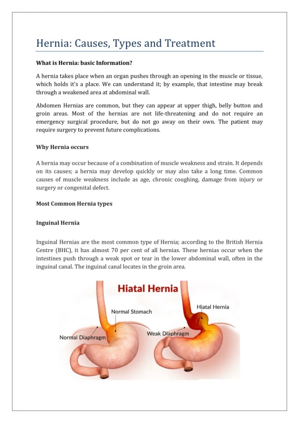
Diaphragmatic/hiatal hernia • It is a commonly caused by automobile-related trauma in small animals although congenital defects of the diaphragm may also result in herniation (eg, peritoneopericardial hernia). • Diaphragmatic hernias are rare in cattle and other species. In swine this occurs sporadically, often in a single animal. They are usually congenital or the result of traumatic accidents. • Dyspnea is usually associated in the acute cases. The degree of dyspnea may vary from subclinical to incompatible with life, depending on the amount of herniated viscera. • If the stomach is herniated, it may bloat and the animal may deteriorate rapidly, as in this case. In chronic cases, systemic signs such as weight loss may be more prominent than respiratory signs. • In cattle and water buffalo, diaphragmatic hernias may be associated with traumatic reticulitis and herniation of the reticulum. Careful physical examination, including auscultation and percussion, usually suggests the presence of thoracic disease. The definitive diagnosis is most frequently made from radiographs. • Loss of diaphragmatic contour, abdominal viscera in the thorax, and displacement of viscera from the abdomen may be apparent. Radiographic contrast studies may be necessary to make the diagnosis. Surgical repair of the hernia is the preferred treatment.
The positive contrast computed radiography of Thoraco-abdominal region using Barium meal revealed diaphragmatic hernia reticulum herniated into thoracic cavity. • A sizeable portion of reticulum was located in thoracic cavity (top right). The case was diagnosed as a diaphragmatic hernia.
Trauma hernia • Typically occur due to sow biting the piglet resulting in a hernia through the abdominal wall. • The hernia is only of consequence if intestinal strangulation occurs. • If the conformation is so badly disfigured that it may result in problems in the slaughterhouse, immediate euthanasia is advised.
Diagnosis • Physical examination. Reduction of the bowel contents in the rupture when squeezed back into the abdomen. • Observing signs such as a swollen, painful, firm umbilical sac along with fine needle aspiration of the swelling to differentiate it from an umbilical abscess. • Diagnostic imaging and ultrasonography. • Laboratory work such as white blood cell counts or fluid collection from the mass for a bacterial culture can also be useful detecting infection.
Treatment options • Routine hernias can be repaired in the field; that is for smaller hernias less than 4 cm methods such as the placement of various hernia clamps, cattle elastrator bands, or the injection of irritating solutions into the tissue. • https://www.youtube.com/watch?v=HiZuRkpPICM • Hernia belts are also used, and are helpful in some cases. The problem with all of these methods is their lack of precision. • Surgical repair is the treatment of choice. Surgery can occasionally perform under sedation with local nerve blocks, but general anesthesia is preferred. The umbilicus for example, its associated structures and all infected tissue are removed. If a large abscess is present it may have to be drained before surgery can be done.