Download
1 / 37
370 likes | 567 Views
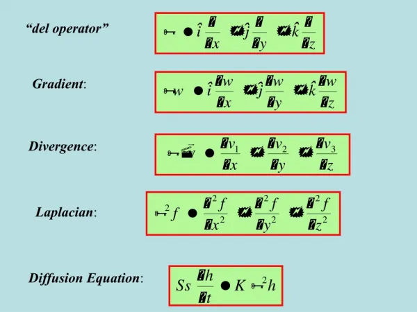
Gradient Echoes, Diffusion, & EPI. Two recent MRI clinical research tools - Echo Planar Imaging ( EPI ) Diffusion Weighted Imaging ( DWI ). Clinical EPI applications. Very rapid imaging (fastest clinical). 128x128 image < 100 ms.

E N D
Gradient Echoes, Diffusion, & EPI • Two recent MRI clinical research tools - • Echo Planar Imaging ( EPI ) • Diffusion Weighted Imaging ( DWI )
Clinical EPI applications • Very rapid imaging (fastest clinical). • 128x128 image < 100 ms. • Multi-slice first pass contrast enhanced brain perfusion. • Functional MRI ( BOLD fMRI ). • Real-time cardiac imaging ?
DWI • Measures diffusion of water. • Diffusion - • Random (Brownian) motion of water. • RMS distance travelled in a fixed time would be a measure of diffusion. Slow diffusion Rapid diffusion
Clinical DWI applications • Ischemia • Ischemic tissue exhibits reduced diffusion. • Intracellular water • - low (restricted) diffusion. • Extracellular water • - higher diffusion. ischemia cytotoxic edema reduction in extracellular volume fraction reduction in overall diffusion
Clinical DWI applications • Ischemic tissue - reduced diffusion. • Reduction observed within about half an hour of ischemia. • T2 increase not seen for 6 - 12 hours (recruitment of excess tissue water). • Assessment of acute stroke. T2 weighted diffusion weighted
Clinical DWI applications • Tumours. • Help distinguish tumour, cystic changes, vasogenic edema, normal white matter. • White matter disease. • Disruption of structure affects local diffusion of water.
S N S N Nuclear Magnetic Resonance Imaging • Nucleus of atom is spinning. • Causes it to behave like a tiny magnet. • Nuclei align (almost) parallel to external magnetic field, like compass needle.
Magnetic Resonance Imaging • Because nucleus is spinning, it can not align exactly parallel to the magnet field. • This causes the N-S axis of the nucleus to rotate around the N-S axis of the main magnetic field. • Precession. North of main magnetic field North of nucleus’s magnetic field
Magnetic Resonance Imaging • The rate of precession is important in MRI. • The number of revolutions per second (frequency) of a precessing nucleus depends on the main magnetic field strength. • Although individual nuclei are not aligned exactly parallel to the main magnetic field, their average alignment is parallel. Millions of nuclei involved in MRI.
Magnetic Resonance Imaging • In the Earth magnetic field • ( 0·00005 T ), hydrogen precesses at about 2100 revolutions per second (Hertz). • In the Vision MRI scanner ( 1·5 T ) hydrogen precesses at 63 million Hertz. • Larmor frequency.
Magnetic Resonance Imaging • “wobble” a nucleus at same rate as it’s precessing tips its alignment away from main magnetic field. • “flip angle”. • Use electromagnetic radiation to do the “wobbling”. • 1·5 T 63 MHz radiowaves (FM) • Interaction due to resonance between precessing nucleus and radiowaves. • Radiowaves in RF pulse. • Applied to whole sample.
Magnetic Resonance Imaging • Switch off RF pulse nuclei realign with main magnetic field. • As they realign, emit radiowaves at Larmor frequency NMR signal out. • NMR signal detected by a RF coil (fancy FM aerial). • Vast majority of NMR signal from hydrogen in water or fat only.
Magnetic Resonance Imaging • MR images are primarily images of water and fat. • To produce an image, apply smaller magnetic fields. • Add / subtract with main magnetic field. • Magnetic field gradients.
1·5 T Magnetic field, B Magnetic field, B 0 x x With gradient Without gradient Magnetic Resonance Imaging • Each point along x axis, different B. • Nuclei precess at different rates. • Emit r/w at different frequencies, depending on their x position. • Spatial encoding of NMR signal.
Free Induction Decay (FID) time NMR signal out RF pulse in Readout gradient time Magnetic Resonance Imaging • To acquire one line of image • Apply gradient during acquisition to spatially encode NMR signal.
Readout gradient time Magnetic Resonance Imaging • Consider nuclei in left & right eyes during FID and 2 ms readout gradient. 1·501 1·5 1·5 1·499 Rt. Lt. Magnetic field, B Magnetic field, B 0 0 x x Freq. 63 63 62·958 63·042 • After 2 ms, Rt. eye: 125916 rotations • Lt. Eye: 126084 rotations • Out of step no gross NMR signal.
Magnetic Resonance Imaging • Practically & theoretically better to separate application of RF pulse and reception on NMR signal. • Arrange peak (in phase) NMR signal in middle of readout gradient. Gradient Echo time RF pulse in NMR signal out Readout gradient time
1·501 1·501 1·499 1·499 Magnetic field, B Magnetic field, B 0 0 x Gradient Echo A B C D Readout gradient 2 2 2 time Rt. Lt. Rt. Lt. x A AB BC C D Rt. eye: 0 126084 125916 252000 377916 Lt. Eye: 0 125916 126084 252000 378084 in step initially de-phased by gradient in step again - echo
1·501 1·501 1·499 1·499 Magnetic field, B Magnetic field, B 0 0 x Gradient Echo - moving nucleus A B C D Readout gradient 2 2 2 time x A AB BC C D Rt. eye: 0 125916 126084 252000 378084 Lt. Eye: 0 126084 125916 252000 377916 moving: 0 126084 126084 252168 378252 Same argument for nuclei only 1 µm apart.
Gradient Echo - moving nucleus • On the scale of an image pixel within the object - • Perfusion: Coherent motion. Entire pixel has same phase shift. • Diffusion: Incoherent random motion. Signal in a pixel is sum of random phase shifts cancel one another out suppression of signal diffusion weighting. • High diffusion large suppression of signal dark pixel in DWI. • Low diffusion little suppression of signal bright in DWI.
Diffusion weighting • Diffusion smaller effect than perfusion small amount of dephasing. • Noticeable DW required very large magnetic gradients. • Separate DW gradients from imaging gradients.
Diffusion weighting RF NMR signal time Readout gradient time Stationary: dephased contributes to nucleus rephased echo. Diffusing: dephased reduced contribution nucleus not fully to echo rephased Diffusion imaging gradients gradients
Diffusion weighting • In practice, use spin echo with DW. 180º NMR signal 90º time Readout gradient time Stationary: dephased rephased contributes to nucleus echo. Diffusing: dephased not fully reduced contribution nucleus rephased to echo Diffusion imaging gradients gradients
Diffusion weighting • Problem: DWI sensitive to any random movements. • Patient movements, e.g., cardiac pulsations, dominate over diffusion. • One solution - use very fast MRI and ‘freeze’ unwanted motions. • Echo Planar Imaging ( EPI )
Gradient echo MRI • Single gradient echo - one line of image. RF time Readout gradient time • Build entire image with FLASH. RF …. time Readout gradient …. time • About 1 second. Poor signal to noise.
EPI • Multiple gradient echoes RF time Readout gradient time Phase: in out in out (stationary nucleus) But, signal still potentially available. Re-phase it with another gradient. Readout gradient time Phase: in out in out in out
EPI • Multiple gradient echoes RF time …. Sign: + - + - + - Readout gradient …. time …. Phase: in out in out in out in out in out in out in out
EPI • NMR signal from one RF pulse lasts between 50 - 200 ms ( T2 decay ). Have to acquire all echoes within this time. • Limited to about 128 echoes, i.e., 128x128 image matrix. • The faster gradients can be switched + to - the more echoes in a fixed time. Entire image in less than 100 ms. Physiological motion frozen. Relatively high S/N (c.f. FLASH). More spatial distortion & artefacts. Not a high resolution technique. High spec. hardware required.
DW-EPI 180º 90º time Readout gradient time Diffusion EP imaging gradients gradients
DW-EPI T2 weighted EPI DW-EPI • High diffusion large suppression of signal dark pixel in DWI. • Low diffusion little suppression of signal bright in DWI.
DW-EPI • DW another MRI parameter • (c.f., T1 and T2 weighting ) • DW-EPI also is heavily T2 weighted (need long TE to fit in extra diffusion gradients). EPI is inherently T2 weighted already. • Bright signal in DWI could also be due to long T2 and vice versa. • “T2 shine through” • To just measure diffusion, calculate the Apparent Diffusion Coefficient ( ADC ). • ‘Apparent’ because averaged over a pixel, and contains some perfusion.
Apparent Diffusion Coefficient No diffusion (stationary) Low diffusion log( DWI ) High diffusion b 0 1000 • b is strength of DW gradients. • Larger b value more DW. • slope of line is the ADC. • To calculate ADC, need minimum of 2 points on line. • We choose b=0 (i.e., T2 weighted EPI) and b=1000 (DW-EPI). • ADC is a quantifiable parameter.
Apparent Diffusion Coefficient b=0 b=1000 ADC map ( T2 weighted ) • In ADC map - • Bright pixel large ADC. • Dark pixel small ADC. • infarct dark • normal brain grey • CSF bright • No potential for “T2 shine through” in ADC map.
Apparent Diffusion Coefficient • Amount of diffusion (ADC) also depends on direction. • In free water, diffusion should be the same in all directions (isotropic) • In structures (e.g., white matter tracts) get more diffusion along the tracts than perpendicular (anisotropic). • Shown DW gradients along x axis. Acquire separate DWI with diffusion along y or z axes. • Construct diffusion tensor. • A tensor gives directional information.
Apparent Diffusion Coefficient Normal brain. ADC map. (amount of diffusion regardless of direction) Relative Anisotropy (RA) map (from tensor). (how uni-directional diffusion is) • White matter tracts bright - • all diffusion in one direction along tracts.
Apparent Diffusion Coefficient Post radiotherapy (AVM). T2 weighted ADC map RA map Stroke. ADC map RA map
Summary • Gradient echo always required to acquire a MR image. • Sensitive to motion. • Can use this to measure diffusion. • Fast MRI sequence needed to freeze all other motions EPI. • Quantify amount of diffusion using ADC maps. • Quantify direction of diffusion using tensor maps. • http://www.nottingham.ac.uk/radiology/