Download
1 / 1
10 likes | 242 Views
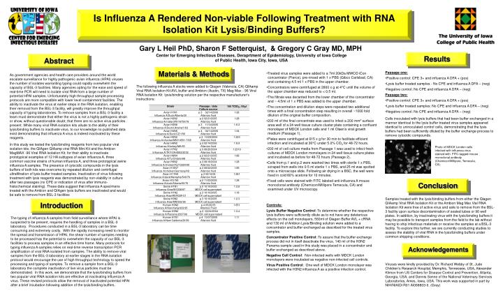
Is Influenza A Rendered Non-viable Following Treatment with RNA Isolation Kit Lysis/Binding Buffers?. Abstract. Acknowledgements. Conclusion. Results. Materials & Methods. Introduction. Gary L Heil PhD, Sharon F Setterquist, & Gregory C Gray MD, MPH.

E N D
Is Influenza A Rendered Non-viable Following Treatment with RNA Isolation Kit Lysis/Binding Buffers? Abstract Acknowledgements Conclusion Results Materials & Methods Introduction Gary L Heil PhD, Sharon F Setterquist, & Gregory C Gray MD, MPH Center for Emerging Infectious Diseases, Deraprtment of Epidemiology, University of Iowa College of Public Health, Iowa City, Iowa, USA • Passage one: • Positive control: CPE 3+ and influenza A DFA + (pos) • Lysis buffer treated samples - No CPE and influenza A DFA – (neg) • Negative control: No CPE and influenza A DFA – (neg) • Passage two: • Positive control: CPE 3+ and influenza A DFA + (pos) • Lysis buffer treated samples: No CPE and influenza A DFA – (neg) • Negative control: No CPE and influenza A DFA – (neg) • Cells inoculated with lysis buffers that had been buffer exchanged in a manner identical to the lysis buffer treated virus samples appeared identical to uninoculated control cells, demonstrating that the lysis buffers had been sufficiently diluted by the buffer exchange process to remove cytocidic compounds. • Treated virus samples were added to a 7ml 20kDa MWCO iCon concentrator (Pierce), pre-rinsed with 1 x PBS (Gibco Carlsbad, CA) and containing 4.5 ml 1 x PBS in the upper chamber. • Concentrators were centrifuged at 2800 x g at 4°C until the volume of the upper chamber was reduced to < 0.5 ml. • The filtrate was decanted from the lower chamber of the concentrator and ~ 4.5ml of 1 x PBS was added to the upper chamber. • The concentration and dilution steps were repeated two additional times with a final concentration step resulting in overall ~1000 fold dilution of the original buffer composition. • 200 ml of the final concentrate was used to infect a 200 mm2 surface area well of a 24 well tissue culture cluster plate containing a confluent monolayer of MDCK London cells and 1 ml Olsen’s viral growth medium (Passage 1). • Plates were centrifuged at 615 x g for 30 min to facilitate efficient infection and incubated at 36°C under 5.0% CO2 for 48-72 hours. • 200 ml of cell culture media from Passage 1 was used to infect fresh cultures of MDCK London monolayers in 24 well tissue culture plates and incubated as before for 48-72 hours (Passage 2). • Cells from p.1 and p.2 were washed two times with sterile 1 x PBS, scraped from wells into 0.5 ml sterile 1 x PBS, and 25 ml was spotted onto a microscope slide. Following air drying in a BSC, the well were fixed in cold 80% acetone for 10 minutes. • Fixed cells were stained with FITC labeled anti-influenza A mouse monoclonal antibody (Chemicon/Millipore Temecula, CA) and examined under UV microscopy. As government agencies and health care providers around the world escalate surveillance for highly pathogenic avian influenza (HPAI) viruses the number of isolates warranting typing could rapidly overwhelm the capacity of BSL-3 facilities. Many agencies opting for the ease and speed of real-time PCR will need to isolate viral RNA from a large number of potential HPAI samples. Unfortunately high throughput sample processing protocols are more compatible with lower level containment facilities. The ability to inactivate the virus at earlier steps in the RNA isolation, enabling their removal from the BSL-3 facility, will greatly improve the throughput capacity of typing laboratories. To remove samples from a BSL-3 facility a team must demonstrate that either the virus is not a highly pathogenic strain or show, without questionable doubt, that there are no active virus particles present. While many viral RNA isolation kits allude to the ability of their lysis/binding buffers to inactivate virus, to our knowledge no published data exist demonstrating that influenza A virus is indeed inactivated by these reagents. In this study we tested the lysis/binding reagents from two popular viral isolation kits, the QIAgen QIAamp viral RNA Mini Kit and the Ambion MagMAX -96 Viral RNA Isolation Kit, for their ability to inactivate prototypical examples of 12 HA subtypes of avian influenza A, three common vaccine strains of human influenza A, and three prototypical swine influenza A samples. The presence of cytocidic compounds in the lysis buffers of both kits was overcome by repeated dilution and centrifugal ultrafiltration of lysis buffer treated samples. Inactivation of virus following treatment with lysis reagents was demonstrated by non-viability in culture after two passages (no CPE or indication of virus after immuno-histochemical staining). These data suggest that influenza A specimens treated with the Ambion and QIAgen lysis buffers are inactivated and would be safe to remove from BSL-3 facilities The following influenza A stocks were added to Qiagen (Valencia, CA) QIAamp Viral RNA Isolation Kit AVL buffer and Ambion (Austin, TX) Mag Max - 96 Viral RNA Isolation Kit lysis/binding solution per the respective manufacturer's instructions: Photo of MDCK London cells infected with influenza virus stained with FITC-tagged mouse monoclonal anitbody (Chemicon/Millipore, Temecula, CA). Samples treated with the lysis/binding buffers from either the Qiagen QIAamp Viral RNA Isolation Kit or the Ambion Mag Max Vial RNA Isolation Kit were free of active virus and safe to remove from the BSL-3 facility upon surface decontamination of sealed tubes or 96 well plates. In addition, by inactivating virus with the lysis/binding buffers it may be possible to transport samples from the field to the lab without having to ship infectious materials or receive the samples at a BSL-3 facility. To explore this further, we are currently conducting studies to assess the stability of viral RNA in the lysis/binding buffers under common shipping conditions. Controls: Lysis Buffer Negative Control: To determine whether the respective lysis buffers were sufficiently dilute as to not have any deleterious effects on the cell monolayers, 560ml of Qiagen Buffer AVL + cRNA and 130 ml of Ambion Lysis/Binding solution were added to a concentrator and buffer exchanged as described for the treated virus samples. Concentrator Positive Control: To assure that the buffer exchange process did not in itself deactivate the virus, 140 ml of the H3N2 Panama sample used in this study was placed in a concentrator and buffer exchanged as described above. Negative Cell Control: Non-infected wells with MDCK London monolayers were incubated as negative non-infected cell controls. Virus Positive Control: One well of MDCK London monolayer was infected with the H3N2 influenza A as a positive infection control. The typing of influenza A samples from field surveillance where HPAI is suspected to be present, requires the handling of samples in a BSL-3 laboratory. Procedures conducted in a BSL-3 laboratory can be time consuming and extremely costly. With the rapidly increasing need to monitor the spread and transmission of HPAI, the shear number of samples needing to be processed has the potential to overwhelm the capacity of our BSL-3 facilities to process samples in an effective time frame. Many protocols for typing influenza A samples relies on real-time reverse transcription PCR amplification of viral RNA isolated from samples. The ability to remove viral samples from the BSL-3 laboratory at earlier stages in the RNA isolation protocol would encourage the use of high throughput technology to speed the processing and typing of samples. To remove a sample from a BSL-3 laboratory the complete inactivation of live virus particles must be demonstrated. In this work, we demonstrate that the lysis/binding buffers from two popular viral RNA isolation kits are effective at inactivating influenza A virus. These revised protocols allow the removal of inactivated potential HPAI after a brief incubation following addition of the lysis/binding buffers. Viruses were kindly provided by Dr. Richard Webby of St. Jude Children’s Research Hospital, Memphis, Tennessee, USA, Alexander Klimov from US Centers for Disease Control and Prevention, Atlanta, Georgia, USA, and Dennis Senne of the National Veterinary Services Laboratories, Ames, Iowa, USA. This work was supported in part by NIH/NIAIDl R01 AI068803-0. (Gray)