Download
1 / 32
400 likes | 866 Views
Principles of instrumentation. Photometry. Photometry means “the measurement of light” If a substance can be converted to a soluble, colored material, its concentration may be determined by the amount of color present in the solution.

E N D
Principles of instrumentation
Photometry • Photometry means “the measurement of light” • If a substance can be converted to a soluble, colored material, its concentration may be determined by the amount of color present in the solution. • Photometer & Spectrophotometer are instruments used for this type of measurement, in which a photocell or photomultiplier tube is used to detect the amount of light that passes through a colored solution from a light source. • The greatest sensitivity is obtained when the light permitted to pass through the solution is of a particular wavelength.(The wavelength shows the maximum absorbance for the solution color).
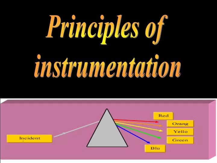
Characteristics of Light • Light is a form of electromagnetic energy that travels in waves. • The wavelength of light is the distance between two beaks. • The light wave, is inversely proportional with its energy. • Resulting in many shades of color.
example: a substance that absorbs violet light at 400 nm reflects all other light and appears as yellow green. The amount of yellow light absorbed varies directly in proportion to the concentration of the blue substances in the solution.
Table-1(wavelengths of various types of Radiation) Energy Wavelength
When the light of an appropriate wavelength strikes a cuvet that contains a colored sample, some of the light is absorbed and the rest is transmitted through the sample to the detector. % percent transmittance which represents the proportion of light reaches the detector. % T = It \ Io x 100 % Where: Io:is the intensity of light striking the sample. It: is the intensity of transmitted light. Beer’s law It Io
Beer’s law • If the concentration of a solution is increased, the It will decrease and then % T is decreased. • The relationship between the concentration and %T is not linear, but if the logarithm of the %T is plotted against the concentration, a straight line is obtained (fig1) -The termabsorbanceis used to represent – log % T A = - log % T = 1/ log % T
Then we can determine the concentration of x substance by measuring both sample and standard absorbance, which can be made by spectrophotometers.
UV – Visible photometry • Typical coloremetric instruments contain five components: • Stable source of radiation energy. • A transparent container for holding the sample. • A device that isolates a restricted region of the spectrum for measurement. • A radiation detector which converts radiant energy to electrical signals. • A signal processor and read out which displays the transudated signals, a meter scale, a digital meter or a recorder chart.
Radiation sources - In UV region: The most commonly used is deuterium lamp or hydrogen lamp. In which a continues spectrum is produced by the excitation of deuterium (D2) or hydrogen at law pressure, and then produced light with (160-375) nm. - In visible region: Tungeston filament lamp is the most commonly used and produces light at (350-2500) nm.
Note: Colorimeters Photometers - Used filters as wavelength selector • Spectrophotometer • - Used monochromators as • Wavelength selector
Sample containers: • Cuvetes that hold the samples must be made of material that passes radiation in the spectral region of interest. • Quartz or fused silica may be used in the spectral region (350-3000 nm), mean it may be used in the UV, visible and a part of infrared. • Silicated glass used in (350- 2000 nm) region. • Plastic is used in the visible region • Radiation detectors and read out.
Hemoglobin Concentration Determination
Hemopoiesis • Is the process of blood cell formation which takes place during the embryonic life in the yolk sac; mesenchymal tissue (liver, spleen, thymus and lymph nodes, bone marrow). • While in late fetus & adult takes place in bone marrow and lymphtic tissues in normal situation (medullary hemopoiesis). • In pathological conditions hemopoiesis is (extramedullary) in the liver, spleen and lymph nodes.
Hemoglobin (Hb) is the oxygen-carrying pigment and predominant protein in the red blood cells. • Hemoglobin is the protein that carries oxygen from the lungs to the tissues and carries carbon dioxide from the tissues back to the lungs. • In order to function most efficiently, hemoglobin needs to bind to oxygen tightly in the oxygen-rich atmosphere of the lungs and be able to release oxygen rapidly in the relatively oxygen-poor environment of the tissues.
Hemoglobin forms an unstable, reversible bond with oxygen. In the oxygenated state it is called oxyhemoglobin and is bright red. • In the reduced state it is called deoxyhemoglobin and is purple-blue. • A hemoglobin molecule consists of four polypeptide chains: two alpha chains, each with 141 amino acids and two beta chains, each with 146 amino acids. • The protein portion of each of these chains is called "globin". • The α and β globin chains are very similar in structure and each one of them is liked with a heme molecule.
A heme group is a flat ring molecule containing carbon, nitrogen and hydrogen atoms, with a single Fe2+ ion at the center. • Without the iron, the ring is called a porphyrin. • Changes in the amino acid sequence of these chains results in abnormal hemoglobin's. • For example, hemoglobin S is found in sickle-cell disease, a severe type of anemia in which the red cells become sickle-shaped when oxygen is in short supply.
polycythemia • Is Above-normal hemoglobin levels • Secondary polycythemia which is may be due to: • Dehydration (sever burns, diarrhea, vomitting, …etc.). • Severe lung or heart disease. • Living at high altitudes. • Heavy smoking. • Primary polycythemia which is due malignant variation in blood cells production in bone marrow.
anemia • Below-normal hemoglobin levels that can be the result of • Iron deficiency or deficiencies in essential vitamins of other elements, such as B12, folate, B6. • Inherited hemoglobin defects, such as sickle cell anemia or Thalassemia. • Other inherited defects affecting the red blood cells. • Excessive bleeding. • Excessive destruction of red blood cells. • Kidney disease. • Bone marrow failure or aplastic anemia. • Cancers that affect the bone marrow.
Reagent:- Cyanmethemoglobin (hemiglobincyanide) (HiCN) reagentcontain potassium cyanide , potassium ferricyanide , dihydrogen potassium phosphate , and a nonionic detergentin 1L of D.W This reagent is pale yellow in color.
Equipments:- Test tubes in rack Automatic pipette with tips Spectrophotometer (540nm)
Specimen:- Whole blood using EDTA as the anticoagulant. Capillary blood may also be used.
Principle:- . 1.Whole Blood is added to cyanmethemoglobin reagent . The potassium ferricyanidein the reagent convert Hb iron from(Fe++)state to (Fe+++)state to form methemoglobin (Hi). 2. Then methemoglobincombine with potassium cyanideto form cyanmethemoglobin (HiCN). 3. Thenonionic detergent improves the lysis of the RBCs and decrease the amount of turbidity resulting from abnormal proteins, such as lipoprotein 4. Absorbance of solution is read in spectrophotometer at 540 nm.
Procedure:- Put 5 ml of pre-prepared working reagent in a test tube . then add 20 µ blood to the test tube . Rinse the pipette 3-5 times with the HiCN reagent until all blood removed from the pipette. 3. Mix well and leave it at room temp. for 3minutes. 4. Measure the absorbance for cyanmethemoglobin at wave length 540 nm against reagent blank.
Calculation • Test Hb concentration = Abs. of test /Abs. of standard * conc. of standard OR • Test Hb concentration = Abs. of test * factor obtained by the standard curve
Discussion:- 1.The dihydrogen potassium phosphatein the reagent in place of sodium bicarbonate to allow the test to be read at the end of 3 min. instead of waiting15 min. with Na bicarbonate. 2. Before the unknown sample is read the solution must be clear if any turbidity is present a falsely elevated result will be obtained. Clouding may be due to:- High WBC count Hemoglobin S &C Lipemic blood 3. Over anticoagulation of the blood does not affect the Hb results
Notes:- Normal values of Hb:- Males14-18 g/dl Females12-16 g/dl Newborns17-22 g/dl This values will vary with:- 1. Age 2. Sex 3. Altitude
*Smokers will have a tendency toward slightly higher Hb levels.