Download
1 / 83
850 likes | 1.07k Views
Focus on Heart Failure. Heart Failure. An abnormal condition involving impaired cardiac pumping/filling Heart is unable to produce an adequate cardiac output (CO) to meet metabolic needs. Heart Failure. Characterized by Ventricular dysfunction Reduced exercise tolerance

E N D
Heart Failure • An abnormal condition involving impaired cardiac pumping/filling • Heart is unable to produce an adequate cardiac output (CO) to meet metabolic needs.
Heart Failure • Characterized by • Ventricular dysfunction • Reduced exercise tolerance • Diminished quality of life • Shortened life expectancy
Heart Failure • Heart failure (HF) is not a disease but a “syndrome.” • Associated with long-standing hypertension, coronary artery disease (CAD), and myocardial infarction (MI)
Heart Failure • Affects about 5 million people in the United States • The most common reason for hospitalization in adults >65 years old
Etiology and Pathophysiology • Primary risk factors • CAD • Advancing age • Contributing risk factors • Hypertension • Diabetes • Tobacco use • Obesity • High serum cholesterol
Etiology and Pathophysiology • Causes of HF may be divided into two subgroups: • Primary • Precipitating • HF is classified as systolic or diastolic failure (or dysfunction).
Etiology and Pathophysiology • Systolic failure • Hallmark finding: Decrease in the left ventricular ejection fraction (EF) • Caused by • Impaired contractile function (e.g., MI) • Increased afterload (e.g., hypertension) • Cardiomyopathy • Mechanical abnormalities (e.g., valve disease)
Etiology and Pathophysiology • Diastolic failure • Impaired ability of the ventricles to relax and fill during diastole, resulting in decreased stroke volume and CO • Diagnosis based on the presence of pulmonary congestion, pulmonary hypertension, ventricular hypertrophy, and normal EF
Etiology and Pathophysiology • Diastolic failure (cont’d) • Caused by • Left ventricular hypertrophy from chronic hypertension • Aortic stenosis • Hypertrophic cardiomyopathy
Etiology and Pathophysiology • Mixed systolic and diastolic failure • Seen in disease states such as dilated cardiomyopathy (DCM) • Poor EFs (<35%) • High pulmonary pressures • Biventricular failure • Both ventricles may be dilated and have poor filling and emptying capacity.
Etiology and Pathophysiology • Compensatory mechanisms are activated to maintain adequate CO. • Sympathetic nervous system (SNS) activation: First and least effective mechanism • Release of catecholamines (epinephrine and norepinephrine) • Increased heart rate (HR) • Increased myocardial contractility • Peripheral vasoconstriction
Etiology and Pathophysiology • Compensatory mechanisms (cont’d) • Sympathetic nervous system (SNS) activation • Over time, these mechanisms are detrimental as they increase the workload of the failing myocardium and the need for O2.
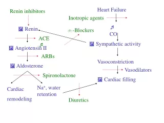
Etiology and Pathophysiology • Compensatory mechanisms (cont’d) • Neurohormonal responses: Kidneys release renin • Renin converts angiotensinogen to angiotensin I. • Angiotensin I is converted to angiotensin II by a converting enzyme made in the lungs.
Etiology and Pathophysiology • Compensatory mechanisms (cont’d) • Neurohormonal responses (cont’d) • Angiotensin II causes • Adrenal cortex to release aldosterone (sodium and water retention) • Increased peripheral vasoconstriction (increases BP) • Response is known as the renin-angiotensin-aldosterone system (RAAS).
Etiology and Pathophysiology • Compensatory mechanisms (cont’d) • Neurohormonal responses (cont’d) • Low CO causes a decrease in cerebral perfusion pressure. • Antidiuretic hormone (ADH) is secreted and causes • Increased water reabsorption in the renal tubules, leading to water retention and increased blood volume
Etiology and Pathophysiology • Compensatory mechanisms (cont’d) • Neurohormonal responses (cont’d) • Endothelin is stimulated by ADH, catecholamines, and angiotensin II, causing • Arterial vasoconstriction • Increase in cardiac contractility • Hypertrophy
Etiology and Pathophysiology • Compensatory mechanisms (cont’d) • Neurohormonal responses (cont’d) • Proinflammatory cytokines (e.g., tumor necrosis factor): Released by cardiac myocytes in response to cardiac injury • Depress cardiac function by causing cardiac hypertrophy, contractile dysfunction, and death of myocytes
Etiology and Pathophysiology • Compensatory mechanisms (cont’d) • Neurohormonal responses (cont’d) • Over time, a systemic inflammatory response is mounted and results in • Cardiac wasting • Muscle myopathy • Fatigue
Etiology and Pathophysiology • Consequences of compensatory mechanisms • Dilation • Enlargement of the chambers of the heart that occurs when pressure in the left ventricle is elevated • Initially an adaptive mechanism • Eventually this mechanism becomes inadequate, and CO decreases.
Dilated & Hypertrophied Heart Chambers Fig. 35-1. A, Dilated heart chambers. B, Hypertrophied heart chambers.
Etiology and Pathophysiology • Consequences of compensatory mechanisms • Hypertrophy • Increase in muscle mass and cardiac wall thickness in response to chronic dilation, resulting in • Poor contractility • Higher O2 needs • Poor coronary artery circulation • Risk for ventricular dysrhythmias
Etiology and Pathophysiology • Counter regulatory processes • Natriuretic peptides: Atrial natriuretic peptide (ANP), b-type natriuretic peptide (BNP) • Released in response to increase in atrial volume and ventricular pressure • Promote venous and arterial vasodilation, reducing preload and afterload • Chronic HF leads to a depletion of these factors.
Etiology and Pathophysiology • Counter regulatory processes (cont’d) • Natriuretic peptides are endothelin and aldosterone antagonists. • Enhance diuresis • Block effects of the RAAS • Natriuretic peptides inhibit the development of cardiac hypertrophy and may have antiinflammatory effects.
Etiology and Pathophysiology • Counter regulatory processes (cont’d) • Nitric oxide (NO) • Released from the vascular endothelium in response to compensatory mechanisms • NO relaxes arterial smooth muscle, resulting in vasodilation and decreased afterload.
Types of Heart Failure • Left-sided HF (most common) from left ventricular dysfunction (e.g., MI hypertension, CAD, cardiomyopathy) • Backup of blood into the left atrium and pulmonary veins • Pulmonary congestion • Edema
Left-Sided Heart Failure Fig. 35-2. Pathophysiology of heart failure. Elevated systemic vascular resistance results in left-sided heart failure that leads to right-sided heart failure. Systemic vascular resistance and preload are exacerbated by the renin-angiotensin-aldosterone system. ADH, Antidiuretic hormone; LA, left atrium; LV, left ventricle; LVEDP, left ventricular end-diastolic pressure; RV, right ventricle.
Types of Heart Failure • Right-sided HF from left-sided HF, cor pulmonale, right ventricular MI • Backup of blood into the right atrium and venous systemic circulation • Jugular venous distention • Hepatomegaly, splenomegaly • Vascular congestion of GI tract • Peripheral edema
Clinical Manifestations: Acute Decompensated Heart Failure (ADHF) • Pulmonary edema, often life-threatening • Early • Increase in the respiratory rate • Decrease in PaO2 • Later • Tachypnea • Respiratory acidemia
Pulmonary Edema Fig. 35-3. As pulmonary edema progresses, it inhibits oxygen and carbon dioxide exchange at the alveolar-capillary interface. A, Normal relationship. B, Increased pulmonary capillary hydrostatic pressure causes fluid to move from the vascular space into the pulmonary interstitial space. C, Lymphatic flow increases in an attempt to pull fluid back into the vascular or lymphatic space. D, Failure of lymphatic flow and worsening of left heart failure result in further movement of fluid into the interstitial space and into the alveoli.
Clinical Manifestations: ADHF • Physical findings • Orthopnea • Dyspnea, tachypnea • Use of accessory muscles • Cyanosis • Cool and clammy skin
Clinical Manifestations: ADHF • Physical findings • Cough with frothy, blood-tinged sputum • Breath sounds: Crackles, wheezes, rhonchi • Tachycardia • Hypotension or hypertension
Clinical Manifestations: Chronic HF • Fatigue • Dyspnea, orthopnea, paroxysmal nocturnal dyspnea • Persistent, dry cough, unrelieved with position change or over-the-counter cough suppressants • Tachycardia
Clinical Manifestations: Chronic HF • Dependent edema • Edema may be pitting in nature • Sudden weight gain of >3 lb (1.4 kg) in 2 days may indicate an exacerbation of HF.
Clinical Manifestations: Chronic HF • Nocturia • Skin • Dusky, cool, damp to touch • Lower extremities: Shiny and swollen, diminished or absent hair growth, pigment changes
Clinical Manifestations: Chronic HF • Restlessness, confusion, decreased memory • Chest pain (angina) • Weight changes • Anorexia, nausea • Fluid retention
Complications: HF • Pleural effusion • Atrial fibrillation (most common dysrhythmia) • Promotes thrombus/embolus formation, increasing risk for stroke • Treatment can include rate control, cardioversion, antidysrhythmics, and/or systemic anticoagulation.
Complications: HF • High risk of fatal dysrhythmias (e.g., sudden cardiac death, ventricular tachycardia) with HF and an EF <35% • HF can lead to severe hepatomegaly, especially with RV failure. • Fibrosis and cirrhosis can develop over time. • Renal insufficiency or failure
Diagnostic Studies • Primary goal: Determine and treat underlying cause • History and physical examination • Chest x-ray • ECG • Lab studies (e.g., cardiac enzymes, BNP)
Diagnostic Studies • Primary goal: Determine and treat underlying cause (cont’d) • Hemodynamic assessment • Echocardiogram • Stress testing • Cardiac catheterization • Ejection fraction
Classification Systems • New York Heart Association Functional Classification of HF • Classes I to IV • ACC/AHA Stages of HF • Stages A to D
Nursing and Collaborative Management • Overall goals of therapy for ADHF and chronic HF • Decrease patient symptoms. • Improve LV function. • Reverse ventricular remodeling. • Improve quality of life. • Decrease mortality and morbidity.
Nursing and Collaborative Management ADHF • High Fowler’s position • Supplemental oxygen • Continuous ECG monitoring • Ultrafiltration: Option for patients with volume overload
Nursing and Collaborative Management ADHF • Circulatory assist devices are used to treat patients with deteriorating HF. • Coexisting psychologic disorders should be addressed.
Nursing and Collaborative Management ADHF • Decrease intravascular volume • Reduces venous return and preload • Loop diuretics (e.g., furosemide [Lasix]) • Ultrafiltration or aquapheresis
Nursing and Collaborative Management ADHF • Decrease venous return (preload) • Reduces the amount of volume returned to the LV during diastole • High-Fowler’s position • IV nitroglycerin
Nursing and Collaborative Management ADHF • Decrease afterload • Improves CO and decreases pulmonary congestion • IV sodium nitroprusside (Nipride) • Morphine sulfate • Nesiritide (Natrecor)
Nursing and Collaborative Management ADHF • Improve gas exchange and oxygenation • Supplemental oxygen • Morphine sulfate • Noninvasive ventilatory support (BiPAP)
Nursing and Collaborative Management ADHF • Improve cardiac function • For patients who do not respond to conventional pharmacotherapy (e.g., diuretics, vasodilators, morphine sulfate) • Inotropic therapy • Digitalis • -Adrenergic agonists (e.g., dopamine) • Phosphodiesterase inhibitors (e.g., milrinone) • Hemodynamic monitoring
Nursing and Collaborative Management ADHF • Reduce anxiety • Distraction, imagery • Sedative medications (e.g., morphine sulfate, benzodiazepines)