Download
1 / 14
180 likes | 484 Views
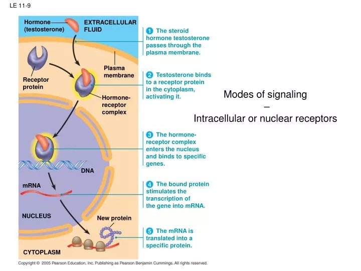
Hormone (testosterone). EXTRACELLULAR FLUID. The steroid hormone testosterone passes through the plasma membrane. Plasma membrane. Testosterone binds to a receptor protein in the cytoplasm, activating it. Receptor protein. Hormone- receptor complex.

E N D
Hormone (testosterone) EXTRACELLULAR FLUID The steroid hormone testosterone passes through the plasma membrane. Plasma membrane Testosterone binds to a receptor protein in the cytoplasm, activating it. Receptor protein Hormone- receptor complex The hormone- receptor complex enters the nucleus and binds to specific genes. DNA The bound protein stimulates the transcription of the gene into mRNA. mRNA NUCLEUS New protein The mRNA is translated into a specific protein. CYTOPLASM LE 11-9 Modes of signaling – Intracellular or nuclear receptors
Cell surface receptors activate a phosphorylation cascade Signaling molecule Receptor Activated relaymolecule Inactiveprotein kinase1 Figure 11.10 Activeprotein kinase1 Inactiveprotein kinase2 ATP Phosphorylation cascade ADP P Activeprotein kinase2 PP P i Inactiveprotein kinase3 ATP ADP P Activeprotein kinase3 PP P i Inactiveprotein ATP P ADP Activeprotein Cellularresponse PP P i
First messenger(signaling moleculesuch as epinephrine) Adenylylcyclase G protein Figure 11.12 GTP G protein-coupledreceptor ATP Second messenger cAMP A signal transduction cascade Proteinkinase A Cellular responses
Signal transduction cascades can be switched on and off Figure 11.11 DG<0 DG<0 Adenylyl cyclase Phosphodiesterase Pyrophosphate H2O P i P ATP cAMP AMP Pyrophosphorylase DG<0 Kinase activity Phosphatase activity ON OFF 2 Pi
Reception Binding of epinephrine to G protein-coupled receptor (1 molecule) Transduction Signal transduction cascades involve signal amplification Inactive G protein Active G protein (102 molecules) Figure 11.16 Inactive adenylyl cyclase Active adenylyl cyclase (102) ATP Cyclic AMP (104) Inactive protein kinase A Active protein kinase A (104) Inactive phosphorylase kinase Active phosphorylase kinase (105) Inactive glycogen phosphorylase Active glycogen phosphorylase (106) Response Glycogen Glucose 1-phosphate (108 molecules)
Second messengers other than cAMP EXTRA-CELLULARFLUID Signaling molecule(first messenger) G protein DAG Figure 11.14-3 GTP G protein-coupledreceptor PIP2 Phospholipase C IP3 (second messenger) IP3-gatedcalcium channel Variousproteinsactivated Cellularresponses Endoplasmicreticulum (ER) Ca2 Ca2(secondmessenger) CYTOSOL
EXTRACELLULARFLUID Plasmamembrane Ca2pump ATP Mitochondrion Figure 11.13 Nucleus CYTOSOL Ca2pump Endoplasmicreticulum(ER) Ca2pump ATP Low [Ca2 ] High [Ca2 ] Key
Intracellular calcium oscillations visualized by fluorescence dye Fura II (a) thymocytes stimulated by extracellular ATP (b) single hepatocyte measured upon epinephrine (adrenaline) stimulation Cox, Lehninger Principles in Biochemistry, chapter 13, figure 18
Growth factor Reception Receptor Cell surface receptors can regulate gene expression, not just nuclear receptors Phosphorylationcascade Figure 11.15 Transduction CYTOPLASM Inactivetranscriptionfactor Activetranscriptionfactor Response P DNA Gene NUCLEUS mRNA
The JAK-STAT signaling pathway. Upon binding ligand, receptor-associated JAKs become activated and mediate phosphorylation of specific receptor tyrosine residues. This leads to the recruitment of specific STATs, which are then also tyrosine-phosphorylated. Activated STATs are released from the receptor, dimerize, translocate to the nucleus, and bind to members of the g-activated site (GAS) family of enhancers. http://www.genome.jp/kegg/pathway/hsa/hsa04630.html J Clin Invest, May 2002, Volume 109, Number 9, 1133-1137
Specificity of signal transduction Signalingmolecule Figure 11.18 Receptor Relay molecules Activationor inhibition Response 2 Response 3 Response 5 Response 4 Response 1 Cell B. Pathway branches,leading to two responses. Cell D. Different receptorleads to a differentresponse. Cell C. Cross-talk occursbetween two pathways. Cell A. Pathway leadsto a single response.
Scaffolding complexes in signal transduction Signalingmolecule Plasmamembrane Figure 11.19 Receptor Threedifferentproteinkinases Scaffoldingprotein
Programmed cell death Ced-9protein (active)inhibits Ced-4activity Ced-9(inactive) Cellformsblebs Figure 11.20/21/22 Death-signalingmolecule Mitochondrion ActiveCed-4 ActiveCed-3 Otherproteases Ced-4 Ced-3 Nucleases Receptorfor death-signalingmolecule Activationcascade Inactive proteins (b) Death signal (a) No death signal