Download
1 / 24
270 likes | 477 Views
OPHTHALMOLOGICAL CHANGES DURING PREGNANCY BY: ABDULKRIM ALKHARASHI. ANATOMY. Accommodation. (A), contraction of ciliary muscles; (B), approximation of ciliary muscles to lens; (C), relaxation of suspensory ligament; (D), increased curvature of anterior surface of lens .

E N D
OPHTHALMOLOGICAL CHANGES DURING PREGNANCY BY:ABDULKRIM ALKHARASHI
Accommodation (A), contraction of ciliary muscles; (B), approximation of ciliary muscles to lens; (C),relaxation of suspensory ligament; (D), increased curvature of anterior surface of lens.
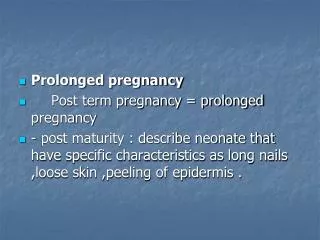
Introduction • The ocular changes that occur in pregnancy are commonly transient in nature but occasionally can be permanent • In addition to the physiological changes in ocular tissues in pregnancy, pathological eye conditions have also been reported
Introduction • While pregnancy can worsen pre-existing ocular conditions such as diabetic retinopathy, it can have beneficial effects in women with glaucoma and uveitis. • Disorders arising in pregnancy, such as preeclampsia and eclampsia, can also present with visual symptoms.
Chloasmaand ptosis • Chloasma, also called the ‘mask of pregnancy’, is a blotchy, brown discoloration that can occur around the eyelids. • It is caused by increase pigmentation related to increased estrogen and progesterone. These changes tend to fade in the postpartum period.
Chloasma and ptosis • Ptosis (drooping of the eyelids) has been reported during and after normal pregnancy1 and is thought to be related to fluid retention and hormonal changes. • It requires no treatment.
Ocular motility defects and Graves’disease • Ocular motility defects can present for the first time during pregnancy • The initial onset of Graves’ disease (exophthalmos)can occur during pregnancy and pre-existing disease can be aggravated early in pregnancy • The treatment requires careful evaluation of the various treatment modalities, as these can pose risks to the fetus
Cornea, Lens andIntraocular pressure • Corneal sensitivity has been found to decrease in most pregnant women and it usually returns to normal by eight weeks postpartum (1) • This can be related to an increase in corneal thickness caused by corneal oedema • 1 - Sunness JS. The pregnant woman’s eye. SurvOphthalmol 1988;32:219–38.
a decrease in tear production occurred during the third trimester of pregnancy in approximately 80% of pregnant women(2) • The curvature of the crystalline lens can increase, causing a myopic shift in refraction • A transient loss of accommodation has been seen during and after pregnancy and in relation to breastfeeding • 2- Imafidon CO, Imafidon JE. Contact lenses in pregnancy. BJOG1992;99:865–7.
Retina and Choroid • The retinal arterioles, venules and capillary bed have been reported as being unchanged during normal pregnancy.(3) • 3-Sunness JS, Santos A. Pregnancy and the Mother’s eye. In: Duane’s Clinical Ophthalmology on CD-ROM. Philadelphia: Lippincott Williams & Wilkins; 2001.
Visual field • There are conflicting reports on changes in the visual field. The reported defects are bitemporal homonymous hemianopiaand central scotoma. • The proposed mechanism is an increase in size of the pituitary gland but only when affecting the optic chiasm.
Visual field bitemporal homonymous hemianopia central scotoma
Disorders of the eye associatedwith pregnancy-related disease • Pre-eclampsia and eclampsia • HELLP syndrome • Occlusive vascular disorders
Pre-eclampsia and eclampsia • Visual disturbances, including scotoma, diplopia, diminished vision and photopsia, are reported in 25% of women with severe pre-eclampsia and in 50% of women with eclampsia • The most common ophthalmological abnormality is retinal arterial spasm and narrowing
Pre-eclampsia and eclampsia • This vascular change is reversible in most women • Other changes associated with retinopathy include haemorrhages, cottonwool spots, retinal oedemaand papilloedema which are seen primarily in women with an underlying chronic systemic disease.
HELLP syndrome • Ocular findings include bilateral serous retinal detachment with yellow/white subretinal opacities and sometimes vitreous haemorrhage.
Occlusive vascular disorders • Pregnancy is associated with a hypercoagulable state and this can affect the retina and choroid • Thrombotic thrombocytopenic purpura (TTP) is rare but can develop in association with pregnancy
Occlusive vascular disorders • Visual symptoms occur in approximately 10% of these women and are generally related to serous retinal detachment, arteriolar constriction and optic disc edema • retinal haemorrhages, exudates, subconjunctivalhaemorrhages, anisocoria (unequal pupils), motility disturbances, ischaemic optic neuropathy, homonymous hemianopia