Download
1 / 85
870 likes | 1.57k Views
DD of parotid lesions. Dr. AHMED REFAEY FRCR RADIOLOGIST. Parotid space. * Paired lateral suprahyoid neck spaces enclosed by superficial layer of deep cervical fascia containing parotid glands, lymph nodes & extracranial CN7 branches. Image gallery.

E N D
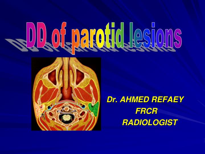
DD of parotid lesions Dr. AHMED REFAEY FRCR RADIOLOGIST
Parotid space * Paired lateral suprahyoid neck spaces enclosed by superficial layer of deep cervical fascia containing parotid glands, lymph nodes & extracranial CN7 branches.
Image gallery • Graphic of a skull base shows PS (green) surrouded by superficial layer, deep cervical fascia(yellowline) . PS abuts stylomastoid foramen (arrow) , mastoid tip (open arrow) & EAC (curved arrow).
Parotid space anatomy • Axial graphic shows superficial layer of deep cervical fascia ( yelow line ) circumscribes PS. • CN7 ( arrow ) divides parotid gland into superficial & deep lobes
Parotid space anatomy • Axial graphic depicts a deep parotid lobe mass pushing the parapharyngeal fat from lateral to medial (arrow) & squeezing through the stylomandibular notch (open arrows)
Image gallery • Sagittal graphic of PS malignancy (arrow) shows typical perineural tumor spread retrograde along CN7 . • Tumor follows CN7 through stylomastoid foramen (open arrow) & up mastoid segment (curved arrow)
Anatomic relationships • Directly medial to parotid space ( PS ) is parapharyngeal space ( PPS ) . • Anterior to PS is masticator space .
Internal structures • Parotid gland -superficial lobe represent about 2/3 of parotid space. -deep lobe projects into lateral PPS • Facial nerve ( CN7) -surgical plane between superficial and deep lobe. • External carotid artery • Retromandibular vein • Lymph nodes • Around 20 lymph nodes found in each parotid gland • Parotid duct -emerges from anterior PS , runs along surface of masseter muscle , arches through buccal space to pierce buccinator muscle at level of upper 2nd molar tooth. • Accessory parotid glands -project over surface of masseter muscle -present in about 20% of normal anatomic dissections.
Key concepts or questions • In mass lesions of PS area, is the mass intra or extraparotid ? -small , intraparotid masses easy to identify. Large , deep lobe masses more troublesome -mass displace PPS medially -stylomandibular notch is widened. • What is mass relationship to facial nerve? -designate mass as superficial , deep or in same plane as intraparotid facial nerve. -superficial lobe mass removed by superficial parotidectomy while deep lobe mass requires total parotidectomy.
. • If malignancy in PS known or suspected ? -T1+C MR should be done to evaluate entire CN7 to root exit zone of CPA , to role if there is evidence of perineural CN7 extension. • Is the PS lesion single or multiple? Unilateral or bilateral ? -multiple bilateral lesions suggest unique DD -Sjogren’s syndrome -BLL-HIV -Warthon tumor -NHl -systemic metastasis
. • Low garde 1ry parotid malignancy may be well circumscribed , hence the surgical rule (( all parotid masses must comeout)). • Facial nerve plane in parotid can only be estimated not seen with imaging. • Parotid LNs are first order drainage for malignancies of adjacent scalp & EAC .
DD of parotid lesions • Congenital • Inflammatory • Neoplasm * benign * malignant - 1ry - 2ry
Congenital *1stbrancheal cleft cyst *infantile hemangioma *lymphangioma Inflammatory *parotiditis *reactive adenopathy *BLL-HIV *Sjogren syndrome *sarcoidosis *Kimura disease Benign tumor *benign mixed tumor *warthin tumor *oncocytoma *facial nerve schwannoma *lipoma Malignant tumor,primary *mucoepidermoid carcinoma *adenoid cystic carcinoma *acinic cell carcinoma *malignant mixed tumor *squamous cell carcinoma Malignant tumor , metastatic *Non-Hodgkin lymphoma *systemic metastasis Differential diagnosis
Parotiditis, acute • Acute infection of parotid gland # bacterial--- acute suppurativeparotitis, usually unilateral, more than 50 years & neonates # viral– acute viral parotitis , more than 75% bilateral , most common cause is mumps, most less than 15 years , peak age 5-9 years. #calculus induced – parotitis2ry to ductal obstruction by stone.
CT findings • NECT • Bacterial and viral – hyperdense enlarged parotid with ill defined margins. • Calculus-induced – parotid duct calculus usually obvious. • CECT • Bacterial – enlarged diffusely enhancing parotid with inflammatory stranding of overlying soft tissues. • Viral – enlarged parotids with mild enhancement. - calculus-induced – parotid duct dilated with enhancing walls.
Image gallery • Axial CECT shows diffusely enlarged and increased in density compared to right side (open arrow)
Image gallery • Axial CECT shows early changes of acute parotiditis. Note subtle asymmetry of parotid density with ill-defined contours and subcutaneous stranding (arrow). Parotid duct is normal ( open arrow)
Image gallery • Axial CECT revealed intraparotid abscess as irregular area of low density (arrow). Note extension of inflammation with carotid space involvement and compressed or thrombosed jugular vein ( open arrow).
Image gallery • Axial CECT shows calculus-induced parotiditis. Note proximal ductal calculus (arrow) with intraglandular ductal radicle enlargement (open arrow). The parotid is enlarged & enhancing without abscess.
Benign lymphoepithelial lesions-HIV( BLL-HIV) • Mixed cystic and solid bilateral intraparotid lesions found in HIV +ve patients. • Best diagnostic clue : multiple cystic and solid masses enlarging both parotid glands usually associated with tonsilar hyperplasia & cervical reactive adenopathy. • Thin rim enhacement of cystic lesions with heterogenous enhancement of solid lesions. • 5% of HIV+ve patients develop BLL of parotids.
BLL-HIV • Axial graphic shows classic findings of BLL-HIV as bilateral intraparotid cysts mixed with bilateral solid lymphoid aggregates (arrows). Note associated adenoidal hypertrophy (open arrows)
BLL-HIV • Axial CECT at level of soft palate shows benign lymphoepithelial lesion of HIV as hypodense cystic & mixed cystic-solid lesions of both parotid glands with thin peripheral enhancement.
Image gallery • Axial CECT reveals bilateral parotid enlargement 2ry to lymphoepithelial lesions of HIV. Note both cystic (arrows) and solid (open arrows) lesions bilaterally affecting the parotid glands.
Image gallery • Axial STIR MR shows bilateral intraparotid hyperintense cystic lymphoepithelial lesions of HIV. Notice both superficial and deep lobes are involved. Arrows marks associated reactive occiptal nodes.
Image gallery • Axial T1 +C MR shows bilateral cystic and solid intraparotid lesions of HIV. Palatine (faucial) tonsils (arrows) are hyperplastic and associated with reactive lateral retropharyngeal nodes (open arrows)
Sjogren’s syndrome • SJS – chronic systemic autoimmune exocrinopathy that causes salivary and lacrimal gland tissue destruction. * 1ry SJS – dry eyes , dry mouth , no collagen vascular disease. * 2ry SJS – dry eyes , dry mouth , with collagen vascular disease, most commonly rheumatoid arthritis.
. • Best diagnostic clue: CT shows bilateral enlarged parotids with multiple cystic and solid intraparotid lesions with or without intraglandular calcification. • Imaging appearence : * early stage – parotids may appear normal * intermediate stage – miliary pattern of small cysts diffusely throughout both glands. * late stage – larger cystic and solid masses in both parotids.
Sjogren syndrome • Axial CECT reveals classic imaging findings of later stage sjogren syndrome with bilateral enlargement, heterogeneity & increased CT density of parotid glands. Note punctate calcifications.
Sjogren syndrome • Axial STIR MR demonstrates early stage MR imaging findings of Sjogren syndrome as bilateral parotid enlargement with miliary diffuse high signal cystic intraparotid lesions.
Image gallery • Axial T1W MR shows multiple low signal cystic lesions involving both parotid glands diffusely. This “ miliary pattern” of diffuse involvement is seen in early stages of Sjogren syndrome.
Benign mixed tumor BMT( pleomorphic adenoma ) • Most common benign parotid space tumor- 80% • Age: most common above 40 y. • Size– variable, may grow to 6-8 cm when in deep lobe. • Large , asymptomatic mass arising from deep lobe of parotid is almost always BMT • 80-90 % of parotid BMT involve superficial lobe. • Multicentric BMT rare ( less than 1%) , but recurrent BMT typically from incomplete resection tends to be multifocal.
Best diagnostic clue: * small BMT– sharply marginated, intraparotid ovoid mass with uniform parenchymal enhancement. * large BMT– more than 2 cm , lobulated mass with inhomogenous enhancement representing foci of necrosis and old hemorrhage. * deep lobe BMT– pear- shaped , inhomogenous mass pushing parapharyngeal space medially.
BMT • Axial graphic depicts a small predominently superficial lobe BMT.
BMT • Axial T1W MR shows small , superficial lobe BMT (arrow). Low signal compared to surrounding parotid is typical. Lateral margin of retromandibular vein (open arrow) marks CN7 plane.
Image gallery • Axial garphic reveals a pear-shaped BMT of the deep lobe of the parotid gland. Notice that despite the size of this tumor, the parapharyngeal fat can still be seen (arrow) being pushed superomedially.
Image gallery • Axial T1+C MR with fat-saturation shows a large , pear-shaped BMT extending from the deep lobe anteromedially. Notice the lesion has pushed the right tonsil into the high oral cavity (arrow)
Image gallery • Axial T1+C MR shows a left parotid tail intermediate sized BMT with inhomogenous enhancement (arrow). As these lesions enlarge , their signal tends to become more inhomogenous on all MR sequences.
Image gallery • Axial CECT shows recurrent BMT as multiple lesions resulting from intraoperative spillage of tumor cells. A larger deep (arrow) & 2 smaller superficial (open arrow) recuurent BMTs can be seen.
Warthin tumor • Benign parotid tumor, sharply marginated ,parotid tail mass with stricking parenchymal inhomogeneity. • Location– most comonly within parotid tail superficial to angle of mandible. • Size– 2-4 cm • Morphology – round to ovoid, well-circumscribed,encapsulated mass or masses ( 20% ). • Parenchymal inhomogeneity is characterestic. • Cystic component in 30% with thin, uniform walls & CT density of 10-20 HU , with minimal enhancement of solid component.
General features • 2nd most common benign parotid tumor. • 20% multicentric, unilateral or bilateral. • Mass is painless, slowly growing. • 90% of patients are smokers. • Increase incidence with radiation exposure • Age– mean age = 60 years.
Image interpretation pearls • Be sure to carefully examine for multiplicity and bilaterality. • Well-circumscribed heterogenous multiple or bilateral parotid masses in asymptomatic patient should be considered warthin tumor.