Download
1 / 1
10 likes | 168 Views
A new "Molecular Scanner" design for interfacing gel electrophoresis with MALDI-TOF. -. +. ThP30 672. Stephen J. Hattan ; Kenneth C. Parker; Marvin L. Vestal SimulTof Corporation, Sudbury , MA. Overlay of averaged s pectra f or all 8 data points at the 3 different

E N D
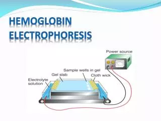
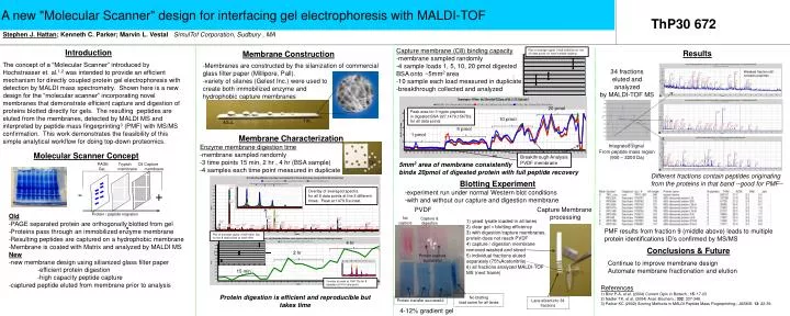
A new "Molecular Scanner" design for interfacing gel electrophoresis with MALDI-TOF - + ThP30 672 Stephen J. Hattan; Kenneth C. Parker; Marvin L. VestalSimulTofCorporation, Sudbury , MA Overlay of averaged spectra for all 8 data points at the 3 different times. Peak at 1479 Da inset. Capture membrane (C8) binding capacity -membrane sampled randomly -4 sample loads 1, 5, 10, 20 pmoldigested BSA onto ~5mm2 area -10 sample each load measured in duplicate -breakthrough collected and analyzed Plot of average signal (1045-3200Da) for the 20 data points for each sample loading Introduction Results Membrane Construction The concept of a “Molecular Scanner” introduced by Hochstrasser et. al.1,2 was intended to provide an efficient mechanism for directly coupled protein gel electrophoresis with detection by MALDI mass spectrometry. Shown here is a new design for the “molecular scanner” incorporating novel membranes that demonstrate efficient capture and digestion of proteins blotted directly for gels. The resulting peptides are eluted from the membranes, detected by MALDI MS and interpreted by peptide mass fingerprinting3 (PMF) with MS/MS confirmation. This work demonstrates the feasibility of this simple analytical workflow for doing top-down proteomics. -Membranes are constructed by the silanizationof commercial glass filter paper (Millipore, Pall). -variety of silanes (Gelest Inc.) were used to create both immobilized enzyme and hydrophobic capture membranes 34 fractions eluted and analyzed by MALDI-TOF MS Weakest fraction still contains peptides 20 pmol Plot of average signal (1045-3200 Da) for the 8 data points at each time Peak area for 3 tryptic peptides in digested BSA 927,1479,1567Da for all data points 10 pmol 1uL 40uL 4 hr 5pmol 1 pmol Membrane Characterization 2 hr Integrated signal From peptide mass region (900 – 3200 Da) Enzyme membrane digestion time -membrane sampled randomly -3 time points 15 min, 2 hr , 4 hr (BSA sample) -4 samples each time point measured in duplicate PAGE TrypsinC8 Capture Gel membrane membrane Molecular Scanner Concept Breakthrough Analysis PVDF membrane 5mm2 area of membrane consistently binds 20pmol of digested protein with full peptide recovery 15 min Different fractions contain peptides originating from the proteins in that band --good for PMF-- Blotting Experiment -experiment run under normal Western blot conditions -with and without our capture and digestion membrane Overlay of peak at 1567 Da for 8 samples at 4 hr time point Protein / peptide migration PVDF Capture Membrane processing Old -PAGE separated protein are orthogonally blotted from gel -Proteins pass through an immobilized enzyme membrane -Resulting peptides are captured on a hydrophobic membrane -Membrane is coated with Matrix and analyzed by MALDI MS New -new membrane design using silianized glass filter paper -efficient protein digestion -high capacity peptide capture -captured peptide eluted from membrane prior to analysis No capture Capture & digestion 1) yeast lysate loaded in all lanes 2) clear gel = blotting efficiency 3) with digestion/capture membranes, protein does not reach PVDF 4) capture / digestion membrane removed washed and sliced 5) individual fractions eluted separately (75%Acetonitrile) 6) all fractions analyzed MALDI-TOF MS (next frame) PMF results from fraction 9 (middle above) leads to multiple protein identifications ID’s confirmed by MS/MS Conclusions & Future Protein capture successful Continue to improve membrane design Automate membrane fractionation and elution References 1) Binz P-A, et al. (2004) Current Opin in Biotech.; 15: 17-23 2) Nadler TK, et al. (2004) Anal. Biochem.; 332: 337-348. 3) Parker KC (2002) Scoring Methods in MALDI Peptide Mass Fingerprinting.; JASMS:13: 22-39. Protein digestion is efficient and reproducible but takes time No blotting load same for all lanes Protein transfer successful Lane sliced into 34 fractions 4-12% gradient gel