Download
1 / 32
580 likes | 1.77k Views
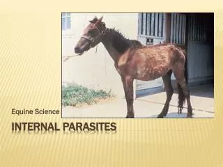
Introduction to Parasites. Introduction. Parasite is an organism baring food and shelter temporarily or permanent and living in or on another organism. The study of parasites is called Parasitology . Parasites can be

E N D
Introduction • Parasite is an organism baring food and shelter temporarily or permanent and living in or on another organism. • The study of parasites is called Parasitology. • Parasites can be • Facultative parasite:parasites able to live both free living and parasite living e.g. Strongyloides species. • Obligate parasite: parasite living permanently in a host and cannot live without a host e.g. Trichomonos species. • Coprozic (spurious) parasites: foreign, pass through alimentally canal without affect. • Clinical Parasitology:deals with animal parasites of man and their medical importance.
Parasitology Divisions of Parasitology: 1. Protozoa 2. Helminthes a. Roundworms (nematodes) b. Flatworms – Cestodes (tapeworm) Trematode (fluke) • Parasitism: organism depend upon another for living, one is living at the expense of the other and harmful, called Parasite, the other organism is called Host.
Host:organism harboring the parasite species may be affected or not. • Classification of Hosts: 1-Definitive host: harbors the adults or final stages or sexual stages (♂♀) in the development of parasite ex: man. 2-Intermediate host: in which you have the larva stages or Inter mediate stages in the development. • Ex: Taenia adult------ man Larva –--- cattle 3-Reservoir host (carrier): the carrier host is well adapted to the parasite and tolerates the infection but serve as source of the infection to other organisms.
Relationships between organisms: • Symbiosis:permanent association between two organisms • Mutualism:two organisms living together, the two organisms benefit. • Commensalism:Two organisms Living together, one is benefited and the other is not been affected. • When the other organism become affected, then the relationship turns = Parasitism. • Zoonosis:disease of animals but can be transmitted to a man. Ex: Hymenolepis nana.
Classification of parasites • General classification: animal parasites are classified according to international code taxonomy – Each parasite belong to a: • Kingdom Phylum Class Order Family Genus Species • Some have further divisions to: Sub – order, super family, sub – species • in classification, scientific parasitic name is of 2 parts: Genus name and species name. Ex: Plasmodium Falciperum Genetic name (one word): plasmodium Species name (two words): plasmodium falciperum. • Genus: means group of close related species. • Species: means population with the same genetic characters.
Entamoeba histolytica • Disease:Amoebiasis Mode of locomotion : Pseudopodia (false feet) • Geo. Dis.: cosmopolitan, but more common in tropical and subtropical countries and in countries with poor sanitation • Habitat: in the lumen of the large intestine (it is pathogenic because it can invade the wall of intestine) • Reservoir: major: humans minor: dogs, pigs, monkeys
Entamoeba histolytica • Morphology: 2 forms Cyst Trophozoite Infective stage: in polluted water and in infected food Pathogenic stage: give pathology as a result of infection
Life cycle: Cyst: infective stage Inters mouth through contaminated food, drink, fly, or through using human stool as fertilizer To L.I. lumen and change into trophozoite (pathogenic stage) Can do erosion through B.V. to liver and other organs Produce lytic enzymes (capable of doing lysis and produce ulcer) Flask shape ulcer
Clinical picture: • Dysentery:blood+mucous diarrhea (as a result of flask shape ulcer wall invasion) • Sever abdominal pain • Tenesmus:sense of incomplete evacuation (the patient at this point should be seeking medical advice) Complication: • intestinal: peritonitis, appendicitis, Hemorrhage • Extra intestinal: Most commonly: liver hepatitis (sever right abdominal pain) Fever amoebic liver abscess (sever pathology in the liver because the inflammation spots came together) shoulder pain and Toxemic manifestations Also in lung, skin, and brain
Plasmodium sp. (Malaria) • Approximately 300 million people worldwide are affected by malaria and between 1 and 1.5 million people die from it every year • Geo. Dis.:Previously extremely widespread, the malaria is now mainly confined to Africa, Asia and Latin America The problems of controlling malaria in these countries are aggravated by inadequate health structures and poor socioeconomic conditions. The situation has become even more complex over the last few years with the increase in resistance to the drugs normally used to combat the parasite that causes the disease. • Causative agent: Malaria is caused by protozoan parasites of the genus Plasmodium. • Species of Plasmodium are: • Plasmodium falciparumthe most widespread and dangerous of the four, if untreated it can lead to fatal cerebral malaria. • Plasmodium vivax. • Plasmodium ovale. • Plasmodium malaria.
Plasmodium sp. (Malaria) • Transmission: Malaria parasites are transmitted from one person to another by the female Anopheles Mosquito. The males do not transmit the disease as they feed only on plant juices. • Reproduction: 1. Sexually reproduction: in anopheles mosquito 2. Asexual reproduction: in human (called sporozoans) in which sporozones multiply to produce merozoites, these, in turn, become trophozoits.
Life cycle: Mosquito bite Plasmodium develops in the gut of mosquito and is passed on in the saliva of an infected insect After 9-16 days they return to the blood and penetrate the red cells, where they multiply again, progressively breaking down the red cells sporozoits are carried by blood to the victim's liver where they form cyst-like structure containing thousands of merozoits This induces bouts of fever and anemia in the infected individual. In cerebral malaria, the infected red cells obstruct the blood vessels in the brain. Other vital organs can also be damaged often leading to the death of the patient.
Plasmodium sp. (Malaria) Pathology and clinical significance: • When merozoits invade the blood cells, using hemoglobin as a nutrient, eventually, the infected red cells rupture, releasing merozoits that can invade other erythrocytes. If a large numbers of red cells rupture at roughly the same time, a paroxysm (sudden onset) of fever can result from the massive release of toxic substance. • Plasmodium falciparum is the most dangerous species. P. malriae, P. vivax, and P. ovale cause milder form of the disease, probably because they invade either young or old red cells, but not both. This is in contrast to P. falciparum, which invades cells of all ages. • Plasmodium falciparum is characterized by persistent high fever and orthostatic hypertension. Infection can lead to capillary obstruction and death if treatment is not introduced.
Toxoplamagondii • Disease is called “Toxoplasmosis” • Geo. Dis.: world wide • Transmission: (1) eating row, undercooked meat of sheep and cow containing viable trophozoits (bradyzoits) (2) swallowing food and water contaminated with infected cat feces (3) Congenital transmission, through placenta (fatal) especially when infection occurs during pregnancy (4) person to person: ex. By blood transfusion or organ transmission • Clinical symptom: -Infection of normal human hosts are common and usually asymptomatic -The infection can be very sever in immunocompromised individuals, who may also suffer recrudescence (relapse) of the infection. -Congenital infections can also be sever, and they are the major cause of blindness in newborns.
What are helminthes? • are a division of eukaryotic parasites that, live inside their host. They are worm-like organisms that live and feed off living hosts, receiving nourishment and protection while disrupting their hosts' nutrient absorption, causing weakness and disease
Nematodes Nematodes are slender, worm-like animals, typically less than 2.5 millimeters long. The smallest nematodes are microscopic, while free-living species can reach as much as 5 centimeters and some parasitic species are larger still.
Ascaris lumbricoides (Roundworm) • Ascaris lumbricoides is the largest nematode (roundworm) parasitizing the human intestine • 1/3 the world population is infected with this worm • Geo.dis.: world wide, common among people with low standard of living and among children • Morphology: Adult: in small intestine adult egg: infective stage
Life cycle: • 2 phases: lung and intestinal • Egg ingested, hatches in duodenum; larvae penetrate intestine wall, enter blood vessels and embolize through liver to lungs. • They then migrate into airspaces, up trachea and are swallowed, taking up permanent adult residence in the small intestine; ~ 2 months from egg to mature adult • Each female produce 200,000 eggs per day • Adult worms can live 1 to 2 years.
Clinical symptoms: • - related to number of worms; small numbers asymptomatic large numbers of adults in intestine – obstruction, pains- at times adults migrate into bile duct, up esophagus or through surgical anast- omoses of intestine- cause malnutrition if in large numbers Lung phase • A.lumbricoides is known as Ascaris pneumonitis. In the lung it causes hemorrhage, inflammation, bacterial infection. It also causes allergy in areas with seasonal trasmission. Typically occurs at 6–14 days after initial exposure. Intestinal phase • The intestinal phase causes malnourishment, intestinal blockage. A.lumbricoides will move around in the body in response to chemotherapy or fever. Typically occurs at 6 to 8 weeks after initial exposure.
Cestodes Tapeworms are ribbon-shaped multi-segmented flatworms that dwell as adults entirely in the human small intestine. The larval forms lodge in skin, liver, muscles, the central nervous system, or any of various other organs
Taeniasaginataor Beef Tape-Worms • habitat is the small intestine—the ileum • Transmission: acquired in humans through the ingestion of raw or poorly cooked meat of infected cows. These cows have been infected via the ingestion of human feces containing the eggs of the parasite • Morphology: Adult is divided into three parts, 1- a head: round and small. It has four suction disks 2- neck: A small, slender neck, about an inch long 3- number of segments. Adult tapeworms and can grow up to 25 meters in the lumen of the intestine, but are usually closer to 5 meters in length Egg present in feces
Taenia saginata Habitat: - adult in small intestine of man only - Egg in feces - larva stage in muscle of thigh, shoulder, neck and heart of cattle only (intermediate host) Clinical symptoms: • High infection: diarrhea and consitipation • Vomiting • Loss of appetite • Anemia Disease is called: Teniasis.
Trematodes Trematodes are flattened oval or worm-like animals, usually no more than a few centimeters in length, although species as small as 1 millimeter and as large as 7 meters are known. Their most distinctive external feature is the presence of two suckers, one close to the mouth, and the other on the underside of the animal.
TrematodesBilharzia (Schistosomiasis) • Disease of the venous system, acquired by people when they come in contact with contaminated water • Adult Schistosomes take up residence in various abdominal veins, depending on the species; they are, therefore called (Blood Flukes) • Very common among children • Geo. Dis.: developing countries, affects up to 200 million people • Transmission:Direct skin penetration Fresh water becomes contaminated by Schistosoma eggs when infected people urinate or defecate in the water. The eggs hatch and the parasites grow and develop inside snails. Schistosoma is not acquired by ingestion of contaminated food, it directly penetrates the skin of swimmers in contaminated rivers and lakes. 2 types of Schistosomiasis: intestinal Schistosomiasis Urinary tract Schistosomiasis
Bilharzia (Schistosomiasis) Pathology: • Intestinal Schistosoma: the primary site of infection is the gastrointestinal tract. Damage to the intestinal wall is caused by the host’s inflammatory response to eggs deposited at that site. The eggs also secret proteolytic enzymes that further damage the tissue. • Urinary tract Schistosoma: The primary site of infection are veins of the urinary bladder, where the organism eggs can induce fibrosis, granulomas, and hematuria • Clinical picture: • Intestinal Schistosoma: GI bleeding, diarrhea, and liver damage. • Urinary tract Schistosoma: hematuria Disease: Bilharzia