Download
1 / 55
560 likes | 743 Views
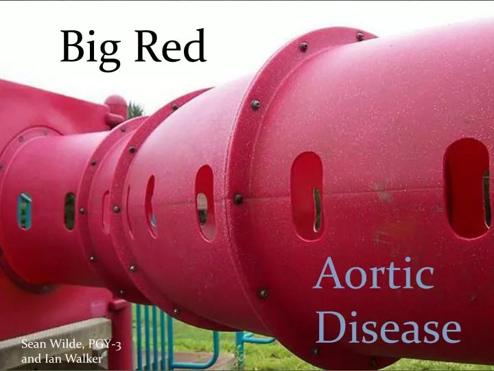
Big Red. Aortic Disease. Sean Wilde, PGY-3 and Ian Walker. Outline. Quick Review Spectrum of Disease Classification Aortic Dissection The proverbial interesting case Avoiding a miss Imaging What’s this D-dimer thing? Emergency Management AAA Diagnosis and management pearls.

E N D
Big Red Aortic Disease Sean Wilde, PGY-3 and Ian Walker
Outline • Quick Review • Spectrum of Disease • Classification • Aortic Dissection • The proverbial interesting case • Avoiding a miss • Imaging • What’s this D-dimer thing? • Emergency Management • AAA • Diagnosis and management pearls
Aneurysms • All 3 layers • TA and AAA • Pseudoaneurysm • No intima, saccular • Trauma, ulcers, infections, surgery • Acute Aortic Syndromes • Thoracic Aortic Dissection • Intramural hematoma • Penetrating atheroscerotic ulcer • Ruptured AAA • Aortocaval fistula • Aortoenteric fistula
From an EM perspective, all variations of acute aortic dissection/hematoma are managed the same:Supportive/Protective therapySurgical consultation
Aortocaval Fistula • Rare • Chest/abdo pain with engorged leg veins • Loud abdominal bruit • Palpable thrill • Needs interventional radiology
Aortoenteric Fistula • Massive upper GI bleed • Fever and back pain • Known AAA or prior AAA repair (especially recent) • No source of bleeding on endoscopy • Sometimes seen on CT scan • Treatment: • Physiologic support • Emergent laparotomy
Case 1 • You are an emerg doc telling a colleague about this crazy case in which you: • A- Totally screwed up and missed the diagnosis of… • B- Actually pulled a rabbit out of your hat and correctly diagnosed… • C- Were barking up the wrong tree when radiology called and said it actually looks like… • And the answer is most often… • Aortic Dissection!
“there is no disease moreconducive to clinical humility than aneurysm of the aorta” Sir William Osler
Aortic Dissection • 2-4 per 100,000 person years • Significant, but much rarer than ACS or PE • On average you will see at least 80 ACS for every dissection. • Classically 50-70 year olds • Does occur in younger populations • M>F • 2-3X more common than ruptured AAA • Mortality estimated 50% if not diagnosed • Mortality increases by 1% every hour of missed diagnosis.
A.D. Risk Factors • Age • 50-70 • Hypertension • Chronic, poorly controlled • Not necessarily acutely • Family history!! • Known prior aortic disease, including repaired • And…
Other AD risk factors • Pregnancy • 3rd trimester highest risk • Bicuspid aortic valve • Connective Tissue disorders • Marfans • Ehlers-Danlos • SLE • Coarctation/congenital heart disease • Turner's syndrome • Cocaine or other stimulant use • Mean 12 hours from last use to presentation • Infectious disease • Syphilis • Endocarditis
Aortic Dissection “Standard of care may be to miss on first presentation.” -Dr. John Eleftriades The great masquerader
Call a lawyer if you miss: • Worst pain of my life • Maximal 10/10 at onset • Ripping, tearing, radiating back or chest pain • Migratory pain • Looks sick • Bilaterally unequal BP and pulse pressure • Wide mediastinum on CHXR
Atypical Presentations are typical… Know them. Chest pain AND leg weakness? My dissection sense is tingling!
Chest Pain AND….. Visual changes Dysphagia, vomiting Limb weakness/ paresthesias Cough Headache Horner’s syndrome Hemiparesis Stroke (Persistent or TIA)
Chest Pain AND….. Paraplegia Lower limb weakness/ paresthesias Anterior cord syndrome Transverse myelitis Progressive myelopathy Spinal Symptoms
Chest Pain AND….. Syncope Painless dissection with syncope is also reported Put AD on your syncope DDx
Chest Pain AND….. Any other unexplained symptom that could be attributable to compromise of an arterial branch, or locally expanding aorta
Unexplained Abdominal Pain AND… • Hypotension • Chest pain • Cocaine use • Historical features (patient or pain) increasing risk of AD • Sick looking patient
Consider aortic dissection any time you have symptoms above AND below the diaphragm
Special Cases: New onset CHF • AD that presents with CHF typically report none to mild pain compared to other dissections • Listen for that murmur of aortic insufficiency • Diastolic aortic murmur is most diagnostic feature • Look for pericardial effusion
Special Cases: ACS • Will see way more ACS than AD • Watch for reg flags on history/physical • Reconsider in MI not responding to ACS therapy • Chest x-ray is very reasonable in every probable MI • Don’t delay ACS treatment to look for dissection except in rare circumstances.
Special Cases: ACS • STEMI • Probably really a STEMI • Dissection is rare, and only 1% of them present as STEMI – Usually Inferior. • But, dissection + lytics • If you’re going to tPA, check: • No history AD red flags • Reasonable CHXR • No loud diastolic murmer
Physical Exam PEARLS • Document bilateral BPs • 10-20% normal people will be different • Many dissections will be normal • Palpable difference in pulse pressures is much more sensitive a finding • Funny looking guy/girl? • Many undiagnosed Marfan’s and other connective tissue disorders • But often will have grossly normal appearance • Check for an aortic murmur • Variable features, but diastolic is classically AI
Aortic Dissection CHXR Top 3 Findings…
Extension beyond intimal calcification
D-Dimer Pros • D-dimer is almost always elevated in aortic dissection • Thrombolysis in false lumen • Majority of studies quote 95-99% sensitivity • Avoid scans especially in young, at risk patients? • Currently lack any definitive negative test outside of major imaging • Search is ongoing for the holy grail of an AD biomarker • Maybe we already have one?
Am J Cardiol 2011;107:1227–1234 - latest D-dimer in AD meta-analysis
D Dimer Cons • Some case reports showing negative dimers in dissections • May not rise in early dissection • May not rise in Intramural hematoma • Typical ED chest pain population not well studied • No validated decision rules for its use • Significance of missing a “small” aortic dissection vs small PE • Will it really decrease scanning? • The PE experience suggests not
D Dimer bottom line? • Probably rules out in most cases, but… • Poorly applicable studies to ED population • Cannot use to derive a true NPV • No valid aortic dissection “low risk criteria” with which to use a D dimer • Multiple potential clinical presentations.
Possible D-dimer roles • Avoid CT in clearly low risk patients • Yet to decide what that actually means • Potentially more useful in the younger population • Maybe Use in conjunction with normal CHXR and low risk history to “reasonably rule out” when you have a better diagnosis. • But you’re going to do way more CT’s doing this • To help distinguish PE/AD from routine MI • Recent small study showing significantly lower elevations in MI vs other conditions. • Trends higher in AD than PE, but was not able to differentiate reliably
Imaging for AD • CT • 94% sens, 90% specific • Gold standard • Want for surgical planning in stable patient • TEE • 97% sens, 75-90% specific • Can do at bedside • EDE ultrasound • Can identify pericardial effusion, sometimes see flap • Cannot use to dx or rule out
Dissection Management:Release the Pressure • Anti-Impulse Therapy! • Lower BP without increasing HR/contractility • Target 100-120 SBP • Start before scanning if highly suspicious
Beta Blocker first (to 60 bpm) • Labetalol • 20mg bolus • 20-80mg q10min, max 300mg • Infusion 0.5-2mg/min • Propranolol • 1-10mg load • 3mg/hr • Esmolol • 500ug/kg bolus • Then infusion (0.1-0.3mg/kg/min) • COPD or labile BP • CCB (diltiazem) in BB intolerant
Then vasodilation to reach goal • Nitroprusside • 0.25-0.5mcg/kg/min • Can use ACEI • Nitroglycerin not recommended (not studied, maybe less effective) • Reasonable if other options impractical (i.e. Rural transport) • Avoid hydralazine, nifedipine • Increases aortic shear wall stress
Hypotense Dissection? • Rule out tamponade • Tamponadevs cardiogenic shock (A. regurg) vshypovolemia • Avoid ionotropes • May need fluid • Some degree of permissive hypotension • Rapid to OR with trauma pack
AAA PEARLS • Vasculopath/smoker • new or different back pain • Syncope and Back pain • Pain to back, flank or groin • Radiation to buttocks, thighs or scrotum • N+V common • Unequal femoral pulses • Sometimes, not always • 50% survival if hypotense
AAA PEARLS Ruptured AAA can present just like renal colic (including hematuria) Renal colic 10-20% time has no hematuria