Download
1 / 28
280 likes | 499 Views
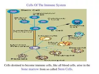
The Cells of the Immune System . Charlie McSharry. Aims and Objectives. By the end of the lectures and lab you should be able to: appreciate the origin of the cells of the immune system from the bone marrow

E N D
The Cells of the Immune System Charlie McSharry
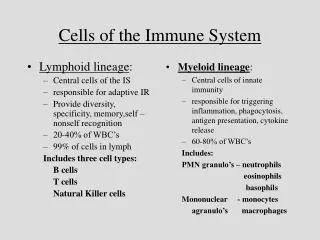
Aims and Objectives By the end of the lectures and lab you should be able to: • appreciate the origin of the cells of the immune system from the bone marrow • describe in simple terms the microscopic appearance of the cells of the immune system: • B and T lymphocytes • plasma cells • macrophages • dendritic cells • describe in simple terms other effector cells such as: • neutrophil, eosinophil and mast cells [polymorphs] • platelets • stromal cells
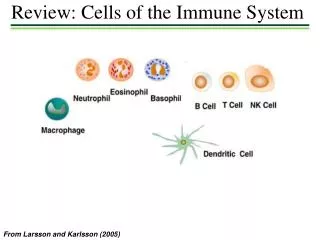
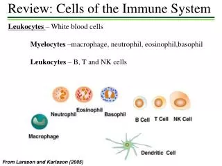
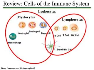
Morphology and staining characteristics of various types of blood cells. Red blood cells and platelets, which both lack nuclei, are the most numerous. Most numerous of the leukocyte populations are the neutrophils. Lymphocytes are the predominant cell type responsible for immune responses.
Granulocytes or Polymorphonuclear (PMN) Leukocytes A group of white blood cells is collectively referred to as granulocytes or polymorphonuclear leukocytes (PMNs). Granulocytes are composed of three cell types identified as neutrophils, eosinophils and basophils, based on their staining characteristics with certain dyes. These cells are predominantly important in the removal of bacteria and parasites from the body. They engulf these foreign bodies and degrade them using their powerful enzymes.
Polymorphonuclear leucocyte -NEUTROPHIL • multi-lobed nucleus. • 50-70% of circulating WBC (higher numbers suggestive of bacterial infection). • The fine granules stain poorly with acidic and basic dyes neutrophil. • Primary granules electron dense - contain bactericidal enzymes • Lysozyme, myeloperoxidase; neutral proteases (i.e. elastase); and acid hydrolases (B-glucoronidase • Secondary granules – smaller, not electron dense. • lysozyme, collagenase and lactoferrin and cathepsin B). • Phagocytosis and killing of ingested microorganisms. • The phagosome fuses with granules to destroy internalized bacteria, oxygen dependent respiratory burst. • DO NOT function as APCs. • Neutrophils are the 1st cells to arrive. A number of substances produced during an inflammatory response recruit neutrophils to a site of inflammation.
LYMPHOCYTES • responsible for the specific immune response. Represent 20-40% of circulating WBC in blood extravasate and enter the tissues – return 99% of cells in lymph • small 6µm, contain a single nucleus, little visible cytoplasm around their nucleus. • T lymphocytes and B lymphocytes and natural killer cells. • T and B lymphocytes are small, motile, nonphagocytic cells which cannot be distinguished from each other morphologically. • Once stimulated with antigen enlarges 15µm into a blast cell. Lymphoblasts further differentiate into effector cells or memory cells. [Plasma cells, T-helper cells, T-cytotoxic cells]. • The memory cells are long-lived cells that reside in the Go phase of the cell cycle until activated by a secondary encounter with antigen. • Different lineages or different maturational stages of lymphocytes can be distinguished by their expression of membrane CD molecules (Cluster of Differentiation (CD)
T and B Lymphocytes • T cells respond to antigens. Some of them (CD4+) secrete lymphokines which act on other cells involved in the immune response. Others (CD8+, cytotoxic) are able to cause lysis of infected cells. • The major function of B lymphocytes is the production of antibodies in response to foreign proteins of bacteria, viruses, and tumor cells. • Antibodies are specialized proteins that specifically recognize and bind to one particular protein that specifically recognize and bind to one particular protein. • Antibody production and binding to a foreign substance or antigen, often is critical as a means of signaling other cells to engulf, kill or remove that substance from the body.
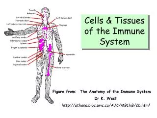
Human lymphoid system • primary organs are bone marrow and thymus • secondary organs and tissues, are lymph nodes and spleen. • These structurally and functionally diverse lymphoid organs and tissues are interconnected by the blood and lymphatic vessels through which lymphocytes circulate. • one bone is shown, but all major bones contain marrow and thus are part of the lymphoid system.
Major lymphoid organs • Thymus. Located in the front of the upper chest, it acts like a nursery for the development of T cells. • Spleen. Located in the upper left side of your abdomen, it filters out foreign organisms that infect your blood, removing old or damaged platelets and red blood cells, storing extra blood and releasing it as needed, and helping form some types of white blood cells. The spleen can be removed if it is damaged, but that may lower your resistance to infection. • Bone marrow. Located in the middle of your bones, most specifically your vertebrae, pelvic, and leg bones, it generates T cells, B cells, and macrophages — cells that travel throughout the body in the blood and tissue fluids. • Lymph nodes. These nodes filter lymph fluid, removing antigens, bacteria, and cancer cells that get trapped in their weblike structure, where macrophages, antibodies, and T cells can destroy them. Hundreds of lymph nodes are located throughout your body, so removing any lymph nodes during breast cancer surgery does not compromise your overall lymph node protection.
Dendritic Cells • originate in the bone marrow • function as antigen presenting cells (APC). • found in the structural compartment of the lymphoid organs • found in the bloodstream and other tissues of the body • capture antigen or bring it to the lymphoid organs where an immune response is initiated.
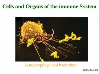
MONOCYTES AND MACROPHAGES • represent 5-8% of WBCs • monocytes enter the tissues through the process of extravastion. • Changes which occur during this transition: Cells enlarge [5-10x] intracellular organelles increase in number and complexity cells acquire increased phagocytic ability increased secretion of many soluble factors • Macrophages play the following important roles: 1) phagocytosis 2) antimicrobial activity 3) secretion of soluble factors • Macrophages are activated by a variety of stimuli in the course of an immune response. - One of the earliest activating signals comes from chemokines. - Phagocyotosis itself is an important activating stimulus. - Macrophages are further activated by cytokines secreted by T helper cells [IFN-gamma] - and by mediators of the inflammatory response - and by various microbial products (such as LPS)
Macrophages • These cells are derived from the bone marrow and have a variety of functions in the immune response: • phagocytosis • secretion of cytokines • The cells performing these various functions have differing microscopic appearances but they are grouped together as the mononuclear phagocytic system.
EOSINOPHIL • (represent 1-3% of circulating WBCs) Possess a bi-lobed nucleus and a heavily granulated cytoplasm. Granules stain orange/red with the acidic dye Eosin Y. Somewhat phagocytic but DO NOT act as APCs. • The major role of the eosinophil is believed to be against parasites, particularly parasitic worms. • Eosinophils kill by ADCC [antibody dependent cell-mediated cytotoxicity] by binding to parasite - specific IgE via cell surface FceRs. • When eosinophils bind to IgE on the surface of a worm, the cell is triggered to degranulate. The contents of the granules cause damage to the worm's tegument. There are many hydrolytic enzymes present in the granules responsible for the anti-helminthic activity. One component which is unique to the eosinophils - and highly toxic to worms - is a substance known as Major Basic Protein (MBP).
BASOPHIL • Only present in the bloodstream, and represent <1% of circulating WBC • Lobed nucleus--more variable, large coarse granules stain blue with basic dye methylene blue. • They play a major role in the allergic response when they release their granules (containing histamine, serotonin, heparin, prostaglandin, etc into the bloodstream following exposure to specific allergens). • Basophils bear Fc receptors for IgE (FceRs) • When an individual is exposed to an allergen, allergen specific IgE is produced. This IgE binds to the surface of basophils [in the sensitization phase of the allergic response]. Upon re-exposure to the allergen, the allergen binds to IgE on the surface of basophils resulting in degranulation [effector phase].
MAST CELLS • Mast cells are released from the bone marrow as undifferentiated precursor cells and do not differentiate until they enter the tissues (skin, connective tissue, mucosal epithelium, etc.) • Morphology and function similar to circulating basophils - but clearly derived from a distinct cell lineage. • Mast cells bear Fc receptors for IgE (FceRs) and contain large numbers of cytoplasmic granules which also play a very important role in the allergic response. • They produce a variety of cytokines • TNF is produced and stored within the cytoplasm of the mast cell, and it can be released quickly following mast cell activation.
Hematopoiesis Regulation of hematopoiesis by cytokines that stimulate the proliferation and/or differentiation of various hematopoietic cells. The bone-marrow stromal cells are the major source of hematopoietic cytokines.
Epidermis Keratinocyte Basement membrane Langerhans cell Melanocyte Dermis Fibroblast Endothelial cell Hypodermis Adipocyte
hepatocyte fibroblast Smooth muscle nerves