Download
1 / 42
420 likes | 538 Views
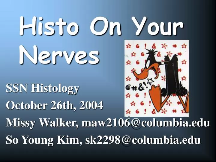
Histo On Your Nerves. SSN Histology October 26th, 2004 Missy Walker, maw2106@columbia.edu So Young Kim, sk2298@columbia.edu. So what’s in a nerve?. Dendrite. Axon Terminal. Perikaryon. Nucleus of myelin-producing cells. Node of Ranvier. Axon. Myelin-Producing Cell. Axon Hillock.

E N D
Histo On Your Nerves SSN Histology October 26th, 2004 Missy Walker, maw2106@columbia.edu So Young Kim, sk2298@columbia.edu
So what’s in a nerve? Dendrite Axon Terminal Perikaryon Nucleus of myelin-producing cells Node of Ranvier Axon Myelin-Producing Cell Axon Hillock
Axon vs. Dendrite NO Synthetic Machinery! Axon Dendrite
Myelin Production in CNS Oligodendrocytes
Spinal Cord Cajal’s Silver
Astrocytes No collagen in the CNS!!!
Somatic Nervous System Innervates skeletal muscle For conscious movement Cell bodies in CNS Autonomic Nervous System Innervates smooth muscle and glands For organ function and regulation Cell bodies in CNS and PNS Sympathetic & Parasympathetic Peripheral Nervous System (PNS)Nervous tissue outside of the brain and spinal cord
Sympathetic GANGLIA in thorax contain: neuronal cell bodies satellite cells (support cells of the neuron) Synapses occur Presynaptic fibers myelinated Postsynaptic fibers unmyelinated ANS - Ganglia
Myenteric plexus in ENS Within Smooth Muscle of Gut NO Collagen (as in CNS) Glial cells (no Schwann cells) Nuclei are euchromatic Gut has “mind of its own” Enteric NS - Ganglia
Organization by Connective Tissues Endoneuriumcollagen fibers associated with individual neurons -Schwann cells within Perineuriumlayer(s) of connective tissue cells, create fascicles Epineurium dense cnxtive tissue binding fascicles into a bundle Cross section of nerve
Epineurium Perineurium Endoneurium Peripheral nerve
Endoneurium Perineurium Epineurium Periph nerve - longitudinal section
Nodes of Ranvier Neurilemma (cytoplasm of Schwann cells) What’s the black? Peripheral Nerve – High Power (Cajal)
What’s the purple? What’s the white? Peripheral Nerve – High Power (H&E)
What’s the purple? Cross Section – Peripheral Nerve
Why is this a Nerve, not a Ganglion? Peripheral Nerve
Key Points for PNS • Ganglia • Contain cell bodies for motor ANS • Satellite cells are the supporting cell bodies (not glial, Schwann, or others of CNS) • Peripheral Nerves • Schwann cells make mylin sheath • Node of Ranvier / saltatory conduction / Na channels • Endoneurium around neurons, which form fascicles • Perineurium around fascicles, which form bundles • Epineurium around bundles
B Questions 1 & 2. . . A
Question 3. . . Figure A Figure B