Download
1 / 20
200 likes | 283 Views
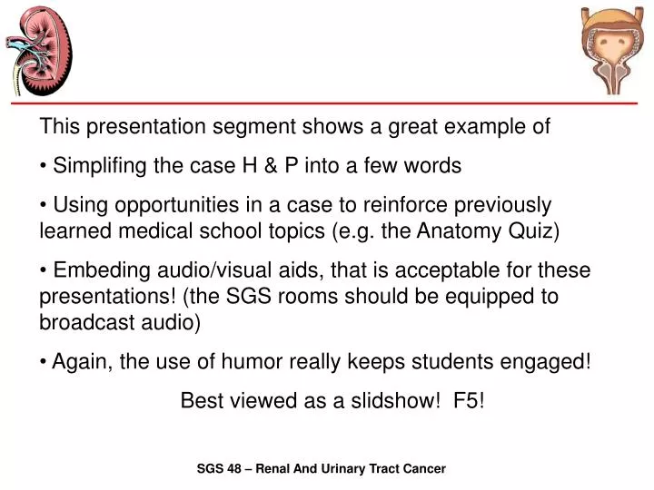
This presentation segment shows a great example of Simplifing the case H & P into a few words Using opportunities in a case to reinforce previously learned medical school topics (e.g. the Anatomy Quiz)

E N D
This presentation segment shows a great example of • Simplifing the case H & P into a few words • Using opportunities in a case to reinforce previously learned medical school topics (e.g. the Anatomy Quiz) • Embeding audio/visual aids, that is acceptable for these presentations! (the SGS rooms should be equipped to broadcast audio) • Again, the use of humor really keeps students engaged! • Best viewed as a slidshow! F5! SGS 48 – Renal And Urinary Tract Cancer
Before We Begin… A Joke: Two old men were arguing the merits of their doctors. The first one said, "I don't trust your fancy doctor. He treated old Jake Waxman for a kidney ailment for nearly a year, and then Jake died of a liver ailment.“ "So what makes you think your doctor is any better?" asked his friend. SGS 48 – Renal And Urinary Tract Cancer
Before We Begin… "Because when my doctor treats you for a kidney ailment, you can be sure you'll die of a kidney ailment." SGS 48 – Renal And Urinary Tract Cancer
Case #2 Renal Cell Carcinoma von Hippel-Lindau SGS 48 – Renal And Urinary Tract Cancer
Case #2 • 32 year old while man • On routine physical exam: • Palpable masses in the left and right flanks. • No pain • Normal urinary and bowel function SGS 48 – Renal And Urinary Tract Cancer
Case #2 Can’t miss the kidneys… Anatomy Quiz Time CT scan of abdomen. Identify major abdominal organs. Identify kidneys and describe abnormalities present. Can a CT scan differentiate between cystic fluid filled structures and solid masses? 18 SGS 48 – Renal And Urinary Tract Cancer
Gas in Colon Hepatic Portal Vein SM Artery and Vein Abd Aorta Transverse / Descending Colon Infer. VC Psoas Major Right Kidney 18 SGS 48 – Renal And Urinary Tract Cancer
Case #2 See multiple, bilateral cysts;variable density (mostly cystic), liver cysts CT scan of abdomen. Identify major abdominal organs. Identify kidneys and describe abnormalities present. Can a CT scan differentiate between cystic fluid filled structures and solid masses? 18 SGS 48 – Renal And Urinary Tract Cancer
Case #2 Renal angiogram. Identify radiographic appearance of neovascularization occurring in renal masses. Correlate areas of neovascularization with abnormalities identified on CT scan. 19 SGS 48 – Renal And Urinary Tract Cancer
Case #2 Neovascularization close to capsule. 19 SGS 48 – Renal And Urinary Tract Cancer
Case #2 - The patient didn’t want a bilateral nephroectomy (hated dialysis). - A kidney transplantation was rejected by the patient (pun intended.) So what to do? Patient underwent an extensive bilateral kidney operation to preserve minimal function without dialysis. SGS 48 – Renal And Urinary Tract Cancer
Case #2 Gross pathologic specimen of the left kidney. Identify normal renal parenchyma and multiple renal masses. Differentiate between solid and cystic lesions within the kidney. Identify renal capsule and relationship of renal masses to the renal capsule and renal fat. 20 SGS 48 – Renal And Urinary Tract Cancer
Case #2 Representative microscopic sections of some of the masses shown in Slide 20. What type of neoplastic cells do you see beneath the cyst lining? Still see clear cells (but not as many), cells layered in sheets. Characteristic of renal cell carcinoma. Proteinacous cyst fluid. 21 SGS 48 – Renal And Urinary Tract Cancer
Case #2 Clear cytoplasms, rich (neo)vascularity Representative microscopic sections of some of the masses shown in Slide 20. What two features characteristic of renal cell carcinoma are seen? 22 SGS 48 – Renal And Urinary Tract Cancer
Case #2 Representative microscopic sections of some of the masses shown in Slide 20. What two features characteristic of renal cell carcinoma are seen? Possible mitosis Obviously something to know about RCC. Clear cytoplasms, rich (neo)vascularity 23 SGS 48 – Renal And Urinary Tract Cancer
G2 G1 G4 G3 Case #2 Renal Cell Carcinoma 4 Fuhrman grades Our patient have RCC, Clear cell carcinoma, nuclear grade I SGS 48 – Renal And Urinary Tract Cancer
Case #2 von Hippel-Lindau loves to spurt other lesions outside the kidney. Where are the most likely sites? Cerebral hemangioblastoma Angiomatosis of the retina Pancreatic cysts and carcinomas Cystadenoma of the epididymis He has this. SGS 48 – Renal And Urinary Tract Cancer
Case #2 Cerebellar hemangioblastoma is a well circumscribed tumor consisting of thin-walled, closely packed capillaries separated by large pale stromal cells. 24 SGS 48 – Renal And Urinary Tract Cancer
Case #2 Cerebellar hemangioblastoma. An immuno-histochemical stainfor factor VIII-related antigen delineates the capillary network. Apparently F8 is a marker for vascular growth. 25 SGS 48 – Renal And Urinary Tract Cancer
Case #2 Summary • What are the anatomic lesions commonly observed in such patients? • Hemangioblastomas in cerebrellum, spinal cord, and retina. • Pancreatic cysts and carcinomas • Cystadenoma of the epididymis • Very rarely pheochromocytoma • What is the vHL genetic characterization and mode of inheritance? • Autosomal dominant, virtually complete penetrance! • Loss of vHL tumor suppressor gene on 3p • Most common RCC etiology (including vHL): Tobacco. • RCC (including vHL) microscopic features: • clear cytoplasm, • neovascularization • hyperchromatic nuclei • pleomorphic nuclei SGS 48 – Renal And Urinary Tract Cancer