Download
1 / 49
500 likes | 566 Views
Tuberculosis: Suspect, Case, Reporting. Acknowledgement. Material in this presentation was assembled from cases managed by Dr. Dana Kissner , Medical Director for TB Control in the Detroit Department of Health and Wellness Promotion, and the staff of the DDHWP TB Program.

E N D
Acknowledgement • Material in this presentation was assembled from cases managed by Dr. Dana Kissner, Medical Director for TB Control in the Detroit Department of Health and Wellness Promotion, and the staff of the DDHWP TB Program. • In partnership with the MDCH TB Control Program, Dr. Kissner has provided this presentation to assist providers in promptly recognizing and diagnosing TB.
Understanding This Presentation • These are real cases that are meant to demonstrate pit-falls in diagnosing TB • The quotes are from the radiologists’ reports • Note that TB is often missed • We hope to improve the diagnosis, initial management, & reporting of • TB suspects • TB cases, laboratory confirmed • TB clinical cases • TB provider diagnosed
Case 137 Year Old AA Man, Cough for 2 Months • Urgent Care – amoxicillin (no chest x-ray) • 6/27/2011 ED Local Hospital • Signed out as “bronchitis & abnormal chest x-ray” • No antibiotic prescribed, thankfully. Why thankfully? • Beware of fluoroquinolones: anti-tuberculosis medications that • Make people with TB feel better. • Can turn TB cultures negative, making it difficult to identify the organism & obtain drug susceptibility • May promote drug resistance if used alone
June 27, 2011 September 1, 2011 “Bilateral hilar enlargement right more than left. Finding could be vascular in nature, however the possibility of neoplastic processes such as lymphoma, or sarcoidosis cannot be completely excluded. Clinical correlation is recommended to determine need for further evaluation by means of a nonemergent enhanced scan of the thorax.” “Enlarged pulmonary hila, unchanged. No active parenchymal disease.” TB?
Who is at risk for exposure to or infection with TB? • Close contacts of person known or suspected to have active TB • Foreign-born persons from areas where TB is common • Persons who visit TB-prevalent countries • Residents & employees of high-risk congregate settings • Health care workers (HCWs) who serve high-risk clients TB skin test 5 mm is +
TB In Michigan 2011 • Cases 170 • Children 6 <4 yrs old • Incidence 1.7 per 100,000 population • Alcohol abuse 14.1% • Injection drug use 4.7% • Non-injection drug 14.1% • HIV + 2.9% • Homeless 10.0% • Black 39.2% • US Born 50.0% TB trends vary considerably throughout Michigan. In 2011, 76.5% of cases were reported from the Detroit metro area.
TB In Detroit 2011* • Cases 53 • Children 4 <22months old • Incidence 7.4 per 100,000 population • Alcohol abuse 30.2% • Injection drug use 15.1% • Non-injection drug 35.8% • HIV + 3.7% • Homeless 28.3% • Black 77.4% • US Born 83.0% * DDHWP jurisdiction Children, adolescents exposed to high risk adults.
Who is at risk for TB after exposure or infection? • Immune suppression • HIV • Organ transplants • Prednisone >15 mg/day > 1 month • TNF-αantagnoists (infliximab, etanercept, adalimumab) • Stable chest x-ray consistent with old healed TB • Children <4 years old • Silicosis, Diabetes, ESRD, underweight, low dose corticosteroids, cancer head & neck TB skin test 5 mm is +
8.8 Million New Cases 128/100,000 38% from India & China India 2.25 million China 1.05 million S Africa 495,000 Indonesia 455,000 Pakistan 405,000
N= 170 Cases >75 % cases in Detroit Metro Area, (Wayne, Oakland, Macomb counties)
Case 2: 55 Year Old Woman • Bi-Polar • Substance abuse • Attends methadone clinic • Fever, chills, cough, sputum X 1 month • Outpatient antibiotics by primary care doctor
Diagnosis Pneumonia: Rx Doxycyline in the ED “New infiltrate with atelectasis is identified in the left lower lobe. Ill-defined parenchymal density is also present in the left upper lobe with a focal central area of lucency which may represent cavitation. Cardiac size is normal. A metallic bullet fragment projects in the right apex. The right lung is clear except for streak atelectasis. Multilevel spurring is present in the dorsal spine. CONCLUSION: Left upper and lower lobe infiltrates with atelectasis.” 3/21/2011
A Prolonged Pneumonia, SepsisFailed Outpatient Treatment “1. Cavitary pneumonia of the left upper lobe. 2. Left lower lobe pneumonia with small pleural effusion. 3. Fibrosis and honeycombing of the right lung apex and base. 4. Bullet in the right posterior thoracic wall” 3/31/2011 Admitted, no isolation, Rx Cefepime & Vancomycin, Erythromycin, Ceftriaxone, “breathing treatments”
Case 3: 46 Year Old Foreign Born* Woman with Multiple Medical Problems • In USA for 20 years • Never-smoker • Diabetes, hypercholesterolemia • Multiple admissions since 2004 • 2004 – Endometriosis, Exploratory lap • 2007 – Cholecystectomy • 2008 – Craniotomy, subarachnoid bleed (other hospital) • Degenerative joint disease, headaches • Cough starting about April, 2011 * High TB Burden Country
CT Scans 6/13, 6/28, 7/5, 8/18 6/13/2011 “2.5 cm rounded pleural based heterogeneous soft tissue mass as discussed. Unless indicated sooner on a clinical basis, recommend 3-month follow-up enhanced chest CT to further characterize. An alternative to obtain in the same added specificity would be to obtain PET/CT imaging at this time, if clinical suspicion for aggressive etiology is present.”
8/18/2011 “Interval development of a new 1.1 cm pleural-based mass in the posteromedial aspect of the superior segment of the right lower lobe. Interval increase in size of the necrotic right pleural-based mass and right subcarinal lymph node. Constellation of findings worrisome for metastatic disease. The need for further evaluation with PET/CT and/or biopsy should be determined clinically”
9/1/2011 • Thoracotomy • Wedge resection RLL “mass” • Wedge resection RUL “mass” • Mediastinal LN sampling • Pathology • Necrotizing granulomas, no AFB • No treatment, no report • 9/2 tissue culture for mycobacteria reported + 10/18 – Health Department notified
Case 4: Health Care WorkerNever Smoker, Cough 5/26/2011 Illustration by Frank Netter, Primary TB
5/26/2011 “Irregular nodular density in the …RML with multiple satellite nodules. There are enlarged lymph nodes in the R hilum & subcarinal region. Findings are suspicious for pneumonia with reactive lymphadenopathy. However possibility of underlying malignancy cannot be excluded completely. Recommended followup with serialchest x rays till complete resolution.”
3 Months Later 8/10/11PET-CT Scan “FDG avid pulmonary nodule in the right middle lobe, along with two FDG avid lymph nodes involving the right hilum and subcarinal region. Findings suspicious for malignancy.”
Panic on 8/18/11 • Wedge resection RML – Large confluent granulomas with extensive central caseating necrosis & groups of AFB • Subsequent sputum negative • Never reported to the Health Department • Discharged on ½ the recommended dose of PZA & EMB
Initial Testing for TB • Collect 3 sputum samples (mycobacteria, AFB) at least 8 hours apart • include at least 1 first morning sample • TB Skin Test or Interferon Gamma Release Assay (IGRA) • HIV test • In-Patient DOT (Nurse watches patient swallow all medications, records doing so)
Laboratory: 6 Essential Tests Ideal Turn-Around Times* • AFB smear: < 24 h • Nucleic acid amplification test**: < 48 h • Growth detection (culture): < 14 days (broth) • TB identification: < 21 days (DNA, broth) • Drug susceptibility testing (DST): < 30 days (directly on broth) – 1o drugs • DST of 2o drugs: < 4 weeks from request or identification of resistant 1o drugs *CDC. Controlling TB in the US…MMWR 2005;54:1-79 **Updated Guidelines for the Use of Nucleic Acid Amplification Tests in the Diagnosis of Tuberculosis MMWR 2009; 58 (01); 7-10
Understanding TB Testing • -AFB smears DO NOT RULE OUT TB!!!!!!!!!!!! • +AFB smears DO NOT DIAGNOSE TB!!!!!!!!!!!!
Next Step • Begin treatment even if sputum AFB smears are negative, based on: • Abnormal chest x-ray (or other x-ray) suggestive of TB • High risk • Clinical symptoms (may be minimal) • + TB skin test or IGRA – Repeat if negative • It can take 10 weeks to become + • - AFB smears DO NOT RULE OUT TB!!!!!!!!!!!!
Therapy for TB • Initial therapy: RIPE* by Directly Observed Therapy (DOT) • Standardized dosing 55 – 75 kg person Add or subtract 1 PZA & 1 EMB if weight is greater or less Rifampin, Isoniazid, Pyrazinamide (PZA), Ethambutol
Therapy for TB • Initial therapy: RIPE* by Directly Observed Therapy (DOT) • Standardized dosing < 55 kg person Add or subtract 1 PZA & 1 EMB if weight is greater or less Rifampin, Isoniazid, Pyrazinamide (PZA), Ethambutol
Therapy for TB • Initial therapy: RIPE* by Directly Observed Therapy (DOT) • Standardized dosing > 75 kg person Add or subtract 1 PZA & 1 EMB if weight is greater or less Rifampin, Isoniazid, Pyrazinamide (PZA), Ethambutol
http://www.umdnj.edu/globaltb/home.htm 1-800-4TB-DOCS
Conditions for Considering the Patient to be Non-Infectious • All of the following are met: • Adequate treatment for 2 weeks or longer • Improved symptoms • 3 consecutive negative sputum smears from sputum collected in 8-24 hour intervals (at least one early morning specimen) • Note: in hospital maintain isolation until all conditions are met
Conditions for Discharge • TB suspect or case has been reported to the local health department • Follow-up plan is in place, home has been evaluated, patient has been interviewed • If possible, the patient is seen in the hospital • DOT arranged, contract signed • Consult your local health department regarding the need for prescriptions
Discharge Planning Checklist MDCH Tuberculosis Hospital Discharge Planning Checklist is available at: www.michigan.gov/tb
Conditions for Discharge • For infectious patients: • The patient has a home to return to • Children < 4 years old & anyone with HIV or other immune compromise are either not in home or have been evaluated & started on “window prophylaxis” (preventive therapy) or treatment for disease • Patient is willing to be on “home isolation”
Health Department • Arranges DOT • Performs contact investigation • Provides expert consultation • Might know where the patient may fit within a cluster • Assures case supervision & completion of Rx
Contacts to The Index Case Family, Friends, Work Place AS1 ♂ >10 Active Prior + Family Health Dep’t 2 TST 1 + Friends Work Place Health Dep’t 1 Health Dep’t 3 TST 1 - TST 1 Pending
What is a Case of TB? • Reporting regions (states, District of Columbia, NY city) report cases to CDC. • Definition of a case • Laboratory confirmed • Clinical Case • Provider Defined
What is a case of TB? • “TB Surveillance. For purposes of surveillance, a case of TB is defined on the basis of laboratory or clinical evidence of active disease due to M. tuberculosis complex.” • “Case classification Confirmed: a case that meets the clinical case definition or is laboratory confirmed.”
Laboratory Confirmed Case • Any 1 of the following is required • 1. Isolation of MTB complex from a clinical specimen • Note: this can be a false + in cases of laboratory or other contamination • 2. Demonstration of MTB complex in a clinical specimen by Nucleic Acid Amplification Test (NAAT) • 3. Demonstration of Acid Fast Bacilli (AFB) from clinical specimen when culture has not or cannot be obtained or is falsely negative or contaminated • The health care worker (Case 4) fit this definition & should have been reported to the health department
Clinical Case. No Lab Confirmation. • Clinical case definition: ALL of the following are present • TST or IGRA is + • Signs or symptoms of TB are present • Abnormal chest x-ray, granulomas on biopsy, lymphocytic pleural effusion, etc.. • Treatment with > 2 anti-TB drugs is started • A diagnostic evaluation is completed
Provider Diagnosis • The criteria for clinical or laboratory case definitions are not met, but a diagnosis of TB is made • Reporting areas have the option of “verifying” a case • Patient remains on treatment and shows improvement • An example may be a lymphocytic pleural effusion with negative TST in a contact to an active case • Another example may be an immunosuppressed patient with signs, symptoms and other clinical evidence of TB
2010: U.S. Cases Without Microbiologic Confirmation • # of Verified Cases 11,182 • + culture 8,413 • + NAA 136 • + AFB smear 72 • Clinical case 1,877 • Provider diagnosis 684 • Percentage • 75% • 1 • 1% • 17% • 6% 2010: 78% cases pulmonary
Latent TB Infection • Cannot be diagnosed while a person remains a TB suspect • Active disease must first be ruled out • Sputum cultures may take up to 6-8 weeks to be positive
Discussion Points • Radiologists are missing cases of TB. • Clinicians are not focusing on risk factors. • Initial investigations are incomplete. • Cases 3 (Foreign Born) & 4 (health care worker) had TST & QFT, respectively. Both were negative. Case 3 was treated with systemic steroids, both probably had early disease. TST / IGRA should be repeated – they can remain negative until 10 weeks after infection. • Failure to involve the health department and report cases is common. • Delayed diagnoses result in poor infection control. • Are we seeing nosocomial spread of TB? • Contacting the health department should result in clear treatment plan for medications, follow-up, contact investigation and infection control.
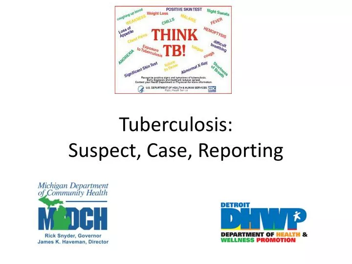
http://www.cdc.gov/tb/publications/Posters/ThinkTB.htm http://www.cdc.gov/tb/statistics/reports/2010/default.htm http://www.umdnj.edu/globaltb/diagnosis&treatment.htm
Resources • Michigan Department of Community Health TB Control Program • www.michigan.gov/tb • Find TB Resources • www.findtbresources.org • CDC Division of TB Elimination • www.cdc.gov/TB/ • NJ Medical School Global TB Institute • www.umdnj.edu/globaltb/home.htm http://www.cdc.gov/tb/education/corecurr/default.htm http://www.cdc.gov/tb/publications/LTBI/default.htm