Download
1 / 30
340 likes | 622 Views
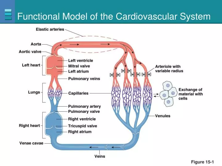
Functional Model of the Cardiovascular System. Figure 15-1. Blood Vessel Structure. Figure 15-2. MIcrocirculation. Capillaries lack smooth muscle and elastic tissue reinforcement which facilitates exchange. Figure 15-3. Elastic Recoil in Arteries. (a). Ventricular contraction.

E N D
Functional Model of the Cardiovascular System Figure 15-1
Blood Vessel Structure Figure 15-2
MIcrocirculation Capillaries lack smooth muscle and elastic tissue reinforcement which facilitates exchange Figure 15-3
Elastic Recoil in Arteries (a) Ventricular contraction Arterioles 1 1 Ventricle contracts. Figure 15-4a, step 1
Elastic Recoil in Arteries (a) Ventricular contraction Arterioles 1 2 1 2 Ventricle contracts. Semilunar valve opens. Figure 15-4a, steps 1–2
Elastic Recoil in Arteries (a) Ventricular contraction Arterioles 1 2 3 1 2 Ventricle contracts. Semilunar valve opens. Aorta and arteries expand and store pressure in elastic walls. 3 Figure 15-4a, steps 1–3
Elastic Recoil in Arteries (b) Ventricular relaxation 1 Isovolumic ventricular relaxation occurs. 1 Figure 15-4b, step 1
Elastic Recoil in Arteries (b) Ventricular relaxation 1 2 Semilunar valve shuts, preventing flow back into ventricle. Isovolumic ventricular relaxation occurs. 1 2 Figure 15-4b, steps 1–2
Elastic Recoil in Arteries (b) Ventricular relaxation 1 2 3 Semilunar valve shuts, preventing flow back into ventricle. Isovolumic ventricular relaxation occurs. 1 2 Elastic recoil of arteries sends blood forward into rest of circulatory system. 3 Figure 15-4b, steps 1–3
Pressure throughout the Systemic Circulation Blood pressure is highest in the arteries and decreases continuously as it flows through the circulatory system Figure 15-5
Blood Pressure • Pulse Pressure = systolic P – diastolic P • Valves ensure one-way flow in veins • MAP = diastolic P + 1/3(systolic P – diastolic P)
Measurement of Arterial Blood Pressure Figure 15-7
Blood Pressure Mean arterial pressure is a function of cardiac output and resistance in the arterioles Figure 15-8
Blood Pressure Blood pressure control involves both the cardiovascular system and the renal system Figure 15-9
Factors that Influence Mean Arterial Pressure Figure 15-10
Arteriolar Resistance • Sympathetic reflexes • Local control of arteriolar resistance • Hormones
Arteriolar Resistance • Myogenic autoregulation • Paracrines • Active hyperemia • Sympathetic control • SNS: norepinephrine • Adrenal medulla: epinephrine
Hyperemia Figure 15-11a
Norepinephrine Tonic control of arteriolar diameter Figure 15-12
Distribution of Blood Distribution of blood in the body at rest Figure 15-13
Blood Flow Blood flow through individual blood vessels is determined by vessel’s resistance to flow Figure 15-14a
Blood Flow Blood flow through individual blood vessels is determined by vessel’s resistance to flow Flow 1/resistance Figure 15-14b
Fluid Exchange at a Capillary Hydrostatic pressure and osmotic pressure regulate bulk flow PLAY Animation: Cardiovascular System: Autoregulation and Capillary Dynamics Figure 15-18a
Blood Pressure KEY Medullary cardiovascular control center Stimulus Sensor/receptor Integrating center Efferent pathway Change in blood pressure Effector Parasympathetic neurons Carotid and aortic baroreceptors Sympathetic neurons SA node Ventricles Veins Arterioles Components of the baroreceptor reflex Figure 15-21
The baroreceptor reflex: the response to increased blood pressure Figure 15-22
The baroreceptor reflex: the response to orthostatic hypotension Figure 15-23
Hypertension The relationship between blood pressure and the risk of developing cardiovascular disease Essential hypertension has no clear cause other than hereditary Figure 15-25
Hypertension • Carotid and aortic baroreceptors adapt • Risk factor for atherosclerosis • Heart muscle hypertrophies • Pulmonary edema • Congestive heart failure • Treatment • Calcium channel blockers, diuretics, beta-blocking drugs, and ACE inhibitors