Download
1 / 159
1.59k likes | 1.6k Views
This article provides an overview of the skeletal system, including its bony and cartilaginous framework, functions, and classification of bones. It also discusses the different parts of the skeletal system, such as bones, joints, cartilages, and ligaments.

E N D
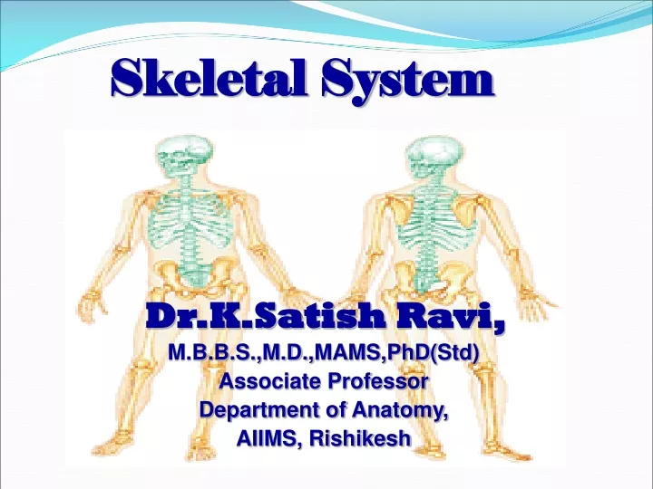
Skeletal System Dr.K.Satish Ravi, M.B.B.S.,M.D.,MAMS,PhD(Std) Associate Professor Department of Anatomy, AIIMS, Rishikesh
SKELETON (skeletos) • Bony & cartilaginous framework of the body • Endoskeleton • Exoskeleton • Functions- • Rigid framework of the body • Protection to the viscera • Provides leverage for body movements.
The Skeletal System • Parts of the skeletal system • Bones (skeleton) • Joints • Cartilages • Ligaments (bone to bone) (tendon=bone to muscle)
Bone :- Highly vascular mineralized connective tissue , consisting of cells & dense intercellular organic matrix impregnated with organic salts. Functions • Shape & Support • Protection • Movement • Storage • Hematopoiesis
CLASSIFICATION of BONES cont’n. • According to location A X I A L skull 22 hyoid 1 ossicles6 vertebrae 26 ribs & sternum 25_ 80
CLASSIFICATION of BONES cont’n. • According to location APPENDICULAR Upper ExtremitiesLower Extremities clavicle 2 hip bone 2 scapulae 2 femur 2 humerus 2 patella 2 radius 2 tibia 2 ulna 2 fibula 2 carpals 16 tarsals 14 metacarpals 10 metatarsals 10 phalanges 28__ phalanges 28__ 64 62
AXIAL SKELETON I. SKULL = skeleton --- head & face = flattened & irregular = united by joints (sutures) • cranium -- skull minus mandible • calvarium -- skull after the bones of the face have been removed • cavities: a. Cranial - contains the brain b. Orbital - contains eyeball & accessory organs c. nasal
Human Skull, Inferior View Figure 5.9
Divisions of the bones of the skull • Cerebral / cranial bones / brain case (8 bones) unpaired (4) paired (4) 1. occipital 1. parietal 2. frontal 2. temporal 3. sphenoid 4. ethmoid b.Facial or visceral cranium paired (12) unpaired (2) a. Nasal a. Vomer b. Lacrimal b. Mandible c. Maxilla d. Zygomatic / malar / cheek bones e. Palatine f. Inferior nasal concha or turbinate
Paranasal Sinuses • Functions of paranasal sinuses • Lighten the skull • Give resonance and amplification to voice
Fontanelle = membrane filled spaces found in the skull of newborn infants e.g.: 1. anterior = largest 2. posterior 3. anterolateral (sphenoidal) 4. posterolateral (mastoid)
AXIAL SKELETON • HYOID BONE = small U-shape; lies in front of the neck = base of the tongue is attached = lies between mandible & thyroid cartilage II. OSSICLES = small bones of the ear a. Stapes (stirrup) 2 b. Incus (anvil) 2 c. Malleus (hammer) 2
AXIAL SKELETON • VERTEBRAL COLUMN = long, curved, slightly movable pillar = united together by cartilage & ligaments = 71 – 75 cm. long = formed by series of bones -- vertebrae FUNCTION: 1.support of the trunk 2. contains & protects the spinal cord & nerves
VERTEBRAL COLUMN Classification of vertebrayoung adult cervical 7 7 thoracic 12 12 lumbar 5 5 sacral 5 1 coccygeal 4 1 33 26 Intervertebral discs = flattened plates of fibrocartilage that are interposed between the adjacent surfaces of the bodies of vertebrae FUNCTION:1. uniting medium between vertebrae 2. main shock absorber 3. give flexibility & movement to the whole vertebral column
VERTEBRAL COLUMN General parts of vertebrae 1. body 5. transverse process 2. arch 6. articular process 3. pedicle or root 7. spinous process 4. lamina 8. spinal or vertebral foramen Special characteristics of individual vertebrae a. Cervical vertebrae (7) = forms the skeleton of the neck, all have transverse foramen atypical cervical vertebrae: 1. atlas -- 1st 2. axis or epistropheus = 2nd 3. 7th cervical vertebrae = spinous process not bifid, small transverse foramen b. Thoracic vertebrae (12) = costal pits - rib attachment = circular vertebral canal
VERTEBRAL COLUMN Special characteristics of individual vertebrae cont’n c. Lumbar vertebrae (5) = presence of mamillary & accessory processes = triangular vertebral foramen d. Sacrum = inverted triangular bone situated between hip bones e. Coccygeal vertebrae (1) = 4 small incomplete vertebrae fused to form the coccyx / tail bone; triangular
AXIAL SKELETON • STERNUM (breast bone) = flat bone, found -- anterior thoracic wall = composed of 2 plates of compact bone with a layer of spongy bone in between containing red bone marrow PARTS: a. Manubrium b. Corpus or body c. Xiphoid process
AXIAL SKELETON • RIBS (12 pairs) = narrow arched flat bones with 2 ends 1. vertebral - posterior; attaches with thoracic 2. sternal - anterior; attaches with costal cartilages Classification of ribs: a. Sternal or true ribs (1st to 7th) - ribs whose costal cartilages are directly attached to sternum b. Asternal or false ribs (8th to 12th) - ribs whose costal cartilages are not attached directly to the sternum but to 7th subdivisions: 1. false rib proper - 8th, 9th, 10th ribs 2. floating or hanging ribs – 11th & 12th
APPENDICULAR SKELETON BONES of the UPPER EXTREMITY (UE) 1. Clavicle (collar bone) 2. Scapula (shoulder blade) – articulates with humerus & clavicle 3. Humerus (arm bone) - longest & largest bone of UE articulates with scapula (above) radius & ulna (below) 4. Radius - lateral bone of the forearm; cup- shaped head 5. Ulna - principal bone of the forearm; longer & larger than radius
APPENDICULAR SKELETON BONES of the UPPER EXTREMITY (UE)cont’n 6. Carpals (wrist bone) - 8 bones arranged into 2 rows - proximal & distal rows She Looks Too Pretty Try To Catch Her Scaphoid, Lunate,Triquetrum, Pisiform,Trapezium, Trapezoid Capitate, Hamate 7. Metacarpals (bones of the hand) - 5 long bones placed between carpals & phalanges - numbered from lateral to medial • Phalanges (bones of the fingers) = 14 long bones of the fingers -- 3 bones except thumb - 2 bones
APPENDICULAR SKELETON BONES of the LOWER EXTREMITY (LE) 1. Hip bone (innominate bone) right & left hip bones + sacrum = pelvic girdle 3 bones: 1. ilium* 2. ischium* 3. pubis* 2. Femur (thigh) = longest, strongest, largest bone in the body 3. Tibia (shin bone) = long bone; anterior, medial, & larger of the 2 bones of the leg 4. Fibula (peroneal bone) = long slender bone placed parallel with the tibia but located laterally *Converge on acetabulum a concave fossa -- articulates with head of femur form hip joint