Download
1 / 28
320 likes | 819 Views
Pemphigus. Primary forms of Pemphigus Deep Pemphigus (above Basal Cell Layer of Epidermis) Pemphigus Vulgaris (most common and most severe) Pemphigus Vegetans (rare Pemphigus Vulgaris variant) Drug-Triggered Pemphigus (unmasks Pemphigus Vulgaris)

E N D
Primary forms of Pemphigus • Deep Pemphigus (above Basal Cell Layer of Epidermis) • Pemphigus Vulgaris (most common and most severe) • Pemphigus Vegetans (rare Pemphigus Vulgaris variant) • Drug-Triggered Pemphigus (unmasks Pemphigus Vulgaris) • Superficial Pemphigus (involves Stratum Granulosum) • Pemphigus Foliaceus • Pemphigus erythematosus (Senear-Usher Syndrome) • Pemphigus herpetiformis
Secondary forms of Pemphigus • Paraneoplastic Pemphigus • Drug-Induced Pemphigus (similar to Pemphigus Foliaceus
PEMPHIGUS VULGARIS • Accounts 70% of all pemphigus • Etiology: • Age: Middle age, rarely affects children • Sex: Both sexes • Races:Affects in eastern countries • Association of other autoimmune disorders Rheumatoid arthritis, Myasthenia gravis, Lupus erythematosus, Pernicious anaemia • Drugs:Penicillamine & Captopril
Uncommon but severe • Potentially fatal autoimmune disorder • Affects skin and mucous membrane. • Two major subtypes: Pemphigus vulgaris Pemphigus foliaceus
·Predisposition to genetic factors: 1. First-degree relatives of patients susceptible to the development of autoimmune diseases 2. Association with HLA-DR4 • Immunological 1. Pemphigusvulgarisantigen:Desmoglein 3 2. Antibody titer: Antibodies of both IgG1 and IgG4 3. Passive transfer:Transplacental transfer of maternal pemphigusvulgaris antibodies may cause transient blisters in neonates. 4.Plasminogen-plasmin system involved in acantholyticprocess:IgGautoantibodies binds with epidermal intercellular material and the adjacent keratinocytes are induced to produced proteolytic enzymes which causes loss of adhesion and intraepidermal split and this process is known as acantholytic process.
Cause • All types of pemphigus are autoimmune diseases in which pathogenic IgG antibodies bind to antigens within the epidermis. The main antigens are desmoglein3 (in pemphigus vulgaris) and desmoglein 1 (in superficial pemphigus). Both are cell-adhesion molecules of the cadherin family , found in desmosomes. The antigen–antibody reaction interferes with adhesion, causing the keratinocytes to fall apart.
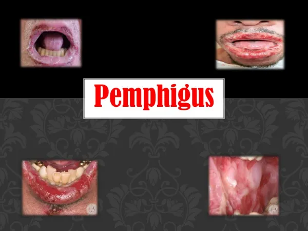
Presentation • Pemphigusvulgaris is characterized by flaccid blisters of the skin and mouth and, after the blisters rupture, by widespread painful erosions. Most patients develop the mouth lesions first. Shearing stresses on normal skin can cause new erosions to form (a positive Nikolsky sign). In the vegetans variant heaped up cauliflower-like weeping areas are present in the groin and body folds. The blisters in pemphigusfoliaceusare so superficial, and rupture so easily, that the clinical picture is dominated more by weeping and crusting erosions than by blisters. In the rarer pemphiguserythematosus, the facial lesions are often pink, dry and scaly.
Associated features and complications • Secondary infection • Extensive lesions associated with water and electrolyte imbalance • Complication of steroids and immunosuppresive drugs.
Mucosal lesions 1.Oral: 50-70% of patients Intact bullae rare in the mouth More commonly: ill-defined, irregularly shaped erosions on buccal, gingiva or palatine 2. Other mucosal surfaces: Conjunctive, Pharynx, esophagus, nose,larynx, urethra, vulva and cervix
Cutaneous lesions: • Localized or generalized Morphology of lesion: Flaccid vesicles on normal skin or an erythematous base---- painful erosion--- heal without scarring -----hyperpigmentation Site: Scalp, face, axillae, groins and pressure points
Nikolsky sign: positive Lesions in skin folds: Vegetating granulations Nail: Dystrophies, acute paronychia, subungual haematomas
Course • The course of all forms of pemphigus is prolonged, even with treatment, and the mortality rate of pemphigus vulgaris is still at least 15%. Superficial pemphigus is less severe. With modern treatments, most patients with pemphigus can live relatively normal lives, with occasional exacerbations.
Pemphigusvegetans: Neumann type & Hallopeau type • A rare variant of pemphigus vulgaris • Characterized by vegetating erosions, primarily in flexures & oral lesions always present • Neumann type: Vesicles & bullae rupture---hypertrophic granulating erosions-- vegetating masses --edges studded with small pustules--erosions at the edge of the lesions induce new vegetation
Hallopeau type: Pustules--vegetating plaques- edges studded with small pustules • Prognosis: Neumann type: Similar to P. vulgaris but prolonged. Hallopeau type: More benign and spontaneous remission
Histology: Spongiosis of basal cells of epidermis followed by suprabasal cleft & acantholytic cells ,basal cells attached to basement membrane Tzank smear: Acantholytic cells
Direct immunofluorescence: IgG in intercellular space of both involved and uninvolved skin (diagnostic) Indirect immunofluorescence: Circulating intercellular antibodies detected in most patients
Differential Diagnosis • Apthous ulcer • Bechet’s syndrome • Epidermolysis bullosa Diagnosis is made by immunofluorescence and histological appearance of bulla.
· • Good oral hygiene • · Potassium permanganate and topical antiseptics • · Candidal infection: topical imidazoles • · Potent topical or intralesional steroids • Prednisolone: prednisolone 1.5mg/kg/day • · Azathioprine 2.5mg/kg/day • · Cyclophosphamide 1-3 mg/kg/day • · Oral or intramuscular gold • · Dapsone • · Plasmapheresis • Treatment
Prognosis · Treatment with systemic steroids reduced mortality 5-15% • Morbidity and mortality • Extent of disease, • Maximum dose of prednisolone required for remission • Presence of other disease • Older patients
Blistering is high in the epidermis, either in the granular layer or just beneath the stratum corneum • Etiology: Accounts 10-20% of pemphigus Antigen: Desmoglein 1, a 160-kDa. • Histopathology: Superficial bullae high in the granular layer or immediately below the stratum corneum Dyskeratotic cells in the granular
Clinical features • Less severe • Onset: Insidious scaly lesions involving the 'seborrhoeic' areas • Scales separate leaving well-demarcated, crusted erosions surrounded by erythema • Erosions are both painful and offensive
PEMPHIGUS ERYTHEMATOSUS • Features of Lupus erythematosus and pemphigus • Oral mucosa rarely involved
Immunological features: · Granular IgG and C3 at BMZ · Intercellular IgG and C3 in the epidermis Circulating antinuclear antibodies • Prognosis: Benign but chronic • Treatment: · Topical or intralesional steroids · Prednisolone 20-40 mg/day
PARANEOPLASTIC PEMPHIGUS • Pemphigus associates with lymphoproliferative disease, thymomas or sarcomas • Clinical features • Overlap with erythema multiforme and lichen planus pemphigoides , Severe mucosal erosions • Polymorphous cutaneous signs • Histopathology: Necrosis of keratinocytes or vacuolar interface dermatitis, Suprabasal clefting
DRUG INDUCED PEMPHIGUS • Drugs may exacerbate or induce pemphigus • Drugs: Thiol- Penicillamine & captopril Non-thiol- enalapril, penicillins, cephalosporins • P. foliaceus and P.erythematosus: common patterns • Circulating auto-antibodies with the same antigenic specificities as in other forms of pemphigus • Prognosis: Recover spontaneously