Download
1 / 25
250 likes | 477 Views
Histology for Pathology Gastrointestinal System and Exocrine Pancreas. Theresa Kristopaitis , MD Associate Professor Director of Mechanisms of Human Disease Kelli A. Hutchens, MD, FCAP Assistant Professor Assistant Director of Mechanisms of Human Disease

E N D
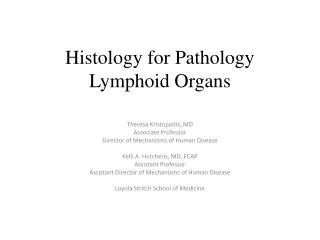
Histology for Pathology Gastrointestinal System and Exocrine Pancreas Theresa Kristopaitis, MD Associate Professor Director of Mechanisms of Human Disease Kelli A. Hutchens, MD, FCAP Assistant Professor Assistant Director of Mechanisms of Human Disease Loyola Stritch School of Medicine
Objectives • On H&E stained sections, identify the four general layers of the digestive tract organs (esophagus, stomach, small bowel, colon): Mucosa; submucosa; muscularis externa, and adventitia/serosa • On H&E stained sections identify the following components of the mucosa: epithelium, lamina propria, muscularis mucosa • Describe the components of the submucosal layer of the digestive organs • Explain the location of Meissner plexus vs Auerbach plexus and describe the function of each • Name the type of epithelium comprising the mucosa of the esophagus, stomach, small bowel, appendix, colon and anal canal. • Identify submucosal glands in the esophagus and describe their function. • Describe the composition of the esophagogastric junction • Name the four parts of the stomach. • Identify gastric pits and explain their function. • On high power H&E stained sections distinguish parietal cells from chief cells. List the substances secreted by each of the cells.
Objectives • Identify the following key components of the small intestine: • Duodenum: villi, Brunner glands • Jejunum: villi, goblet cells • Ileum: villi, goblet cells, Peyer patches • Define Crypts (or Glands) of Lieberkuhn. • Contrast vilivsplicaecirculares • On H&E stained sections distinguish colon from small intestine. • Define taenia coli. • In H&E stained sections of pancreas distinguish the endocrine components of the pancreas from the exocrine components. • In H&E stained sections of pancreas identify pancreatic acinar cells vs ducts.
Oral Cavity • Inner surface of the lips, cheeks, soft palate, surface of tongue, and floor of the mouth • Nonkeratinized stratified squamous epithelium • Lamina propria • Submucosa • Gingiva and hard palate • Keratinized stratified squamous epithelium • Lamina propria • Tongue: specialized mucosa with papillae
Digestive Tract • Comprised of hallow organs • Esophagus • Stomach • Small intestine • Large intestine • Histologic organization: • Mucosa: • Epithelium, lamina propria, muscularis mucosa • Submucosa: • connective tissue, vessels, and Meissners plexuses, some times mucous glands • Muscularis externa: 2-3 layers of smooth muscle (plus skeletal muscle in esophagus), myenteric (Auerbach) plexus in between muscle layers • Serosa and adventitia: Outermost layer of loose connective tissue and blood vessels. Call serosa if covered my mesothelium; adventitia otherwise
Esophagus • Mucosa: non-keratinizing stratified squamous • Submucosa: contains mucous glands • Increased mucous glands at lower esophagus (GE junction) to protect esophagus from gastric juices • Muscularisexterna: inner circular and outer longitudinal • Contains skeletal muscle fibers
Esophagus Squamous mucosa Submucosa Muscularis Externa Mucosa Muscularis mucosa Adventitia / Serosa
Stomach • Anatomically and histologically divided into cardia, fundus body, and pylorus • Cardia: first section; separate from esophagus by cardiac sphincter. Glands contain mucus-secreting cells, stem cells, enterendocrine, and occasional parietal cells • Fundus and body: largest portion. Fundic glands contain parietal cells and chief cells with some stem cells, mucous cells and enteroendocrine cells • Pyloric region: lower end that connects to duodenum; ends at pyloric sphincter. Glands primarily mucus-secreting cells and two special endocrine cells: gastrin-secreting (G cells) and somatostatin-secreting cells (D-cells)
Mucosa of the Stomach Columnar Epithelium Gastric Pit
Fundic Mucosa Parietal Cells : secrete HCL Fundic Gland Chief Cells : secret zymogen granules containing pepsinogen
Small Intestine • Major site for absorption of nutrients and its mucosa is made for this! • Villi and microvilli • Glands (crypts) of Lieberkuhn in lamina propria that open into the mucosa at the base of villi. • Paneth cells with pink granules at the base of the crypts secrete lysozymes, tumor necrosis factor-alpha, and defensins. • Submucosa with Meissner plexus • Three sections • Duodenum: Brunner glands (mucus secreting) • Jejunum: long villi and increased goblet cells – no Brunner glands or Peyer patches • Ileum: Short villi, many goblet cells, and lymphatic nodules in the submucosacalled Peyer patches
Duodenum Mucosa Brunner’s Glands Paneth Cells
Jejunum Goblet Cells
Ileum Paneth Cells Peyers patches
Large Intestine • Major site of absorption of water and salts • Mucosa: No villi, gland of Lieberkuhn with many goblet cells and no paneth cells. • Submucosa: no glands • Muscularis externa: Inner circular muscle plus outer longitudinal muscle with myenteric (Auerbach) plexus between. • Outer longitudinal muscle creates three narrow bands called tenia coli. • Four anatomic sections • Cecum : • Appendix • Colon • Rectum and anal canal
Large Intestine Mucosa Goblet Cells
Colon Wall Muscularis externa (circular portion) Mucosa Submucosa
Appendix Lymphatic Nodules
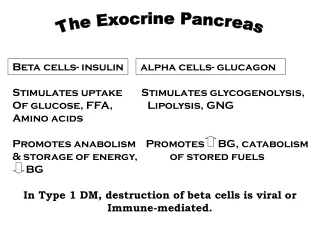
Exocrine Pancreas • Many serous secretory cells (pancreatic acinar cells) form acinar structures. • Cytoplasm contains zymogen granules • Secretions carried by main duct to join bile duct at the ampulla in the duodenum
Main pancreatic duct and surrounding acini Main Duct Pancreatic Acini