Download
1 / 20
200 likes | 299 Views
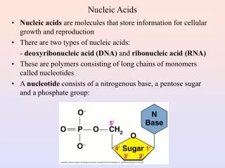
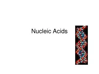
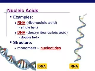
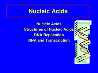
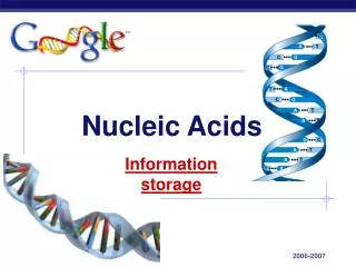
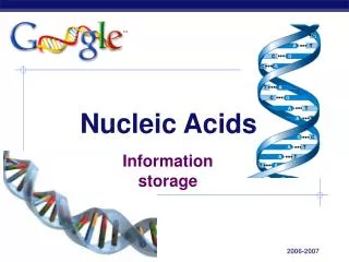
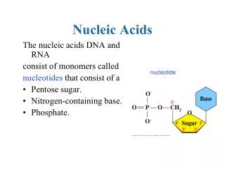
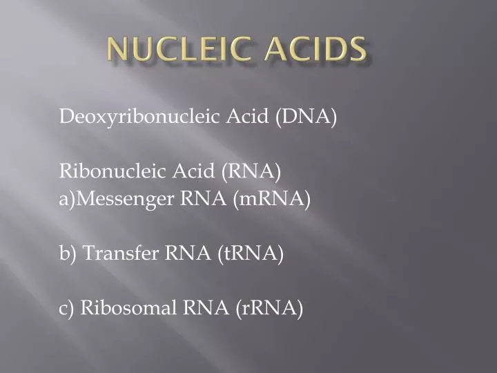
Nucleic Acids. Deoxyribonucleic Acid (DNA) Ribonucleic Acid (RNA) a)Messenger RNA (mRNA) b) Transfer RNA (tRNA) c) Ribosomal RNA (rRNA). DNA. Double helix shape was discovered in 1953 by James Watson and Francis Crick Contains the genetic code for the production of Protein

E N D
Nucleic Acids Deoxyribonucleic Acid (DNA) Ribonucleic Acid (RNA) a)Messenger RNA (mRNA) b) Transfer RNA (tRNA) c) Ribosomal RNA (rRNA)
DNA • Double helix shape was discovered in 1953 by James Watson and Francis Crick • Contains the genetic code for the production of Protein • Polymer composed of Nucleotides • Each DNA triplet codes for one amino acid • The “coding” side is complimentary to the “complimentary” side
DNA is Made of Two Long Chains of Nucleotides Joined by Hydrogen Bonds
chromatin This …. …. not this is what’s found in the cell. DNA is Almost Always Wrapped Around Proteins
Nitrogen Bases • Adenine and Guanine are “Double ringed” structures and are called Purines • Thymine and Cytosine (Uracil) are single ringed structures and are called Pyrimidines
Adenine (Purine) can only bond to Thymine (Pyrimidine) • Guanine can only bond to Cytosine • An exposed Phosphate indicates the 5 prime end • No exposed phosphate indicates the 3 prime end. • DNA Structure • Draw a DNA triplet with the 5 prime on the top left side and the nitrogen base sequence A-C-G on the left side. Remember to include the complimentary side
Complementary Base Pairing Allows Each Strand of DNA to Serve as a Template for DNA Replication Replication always proceeds from the 5’ to the 3’ DNA is a perfect illustration of function following form (structure dictates function).
Replication Enzymes • Helicase – splits the double helix • DNA polymerase – attaches the free-floating nucleotides to the exposed nitrogen bases of the original DNA • RNA primase – inserts short, temporary strands of RNA during replication. • Exonuclease – strips away the RNA strands • Ligase – inserts phosphate groups in any gaps that are left • Replication • Replication Enzymes
DNA Replication – Something Old and Something New In Each Daughter Molecule
RNA • Messenger RNA (mRNA) • - single stranded molecule • - sugar is called Ribose • - thymine is replaced by Uracil • - carries a message from the nucleus to the cytoplasm that determines the amino acid sequence in newly produced protein. • - each group of 3 nitrogen bases is called a codon
Draw a mRNA strand with one codon. Include Uracil as one of the nitrogen bases
RNA • Transfer RNA (tRNA) • - odd shaped molecules with about 75 nucleotides in it’s structure. • - 3 exposed nitrogen bases called anti-codons. • - tRNA molecules carry amino acids to the ribosome during protein synthesis • - each tRNA molecule can only carry one amino acid
RNA • Ribosomal RNA • - main structural component of a ribosome.
A ribosome consists of 2 subunits: 50s sub-unit and a 30s sub-unit compose a 80s ribosome.